Describe the structure and role of microfilaments.
They are linear, thin filaments composed of many actin monomers. Microfilaments are thinner than either microtubules or intermediate filaments.
As actin polymers, microfilaments play a crucial role in muscle contraction and also facilitate cytokinesis.
Key Terms
Describe the structure and role of microfilaments.
They are linear, thin filaments composed of many actin monomers. Microfilaments are thinner than either microtubules or intermediate filaments.
...Describe the structure and role of intermediate filaments.
They are fibers composed of various cell-specific proteins; many are composed of keratin. Intermediate filaments are thicker than microfilaments bu...
Describe the structural role of microtubules.
They are thick, hollow tubulin polymers. Specifically, units of alpha- and beta-tubulin dimerize, and many of these dimers come together to form a ...
What structural and functional differences exist between cilia and flagella?
Cilia are short, found in large numbers, and beat rhythmically in a back-and-forth pattern.
Flagella are longer, with only a...
Describe the composition of the plasma membrane.
It consists of a phospholipid bilayer, with polar heads on the exterior (pointing toward the extracellular fluid and cytoplasm) and nonpolar tails ...
What will happen to a human cell when it is placed in a hypertonic solution?
Water will exit the cell, causing it to shrivel.
A hypertonic solution is one that contains a comparatively high amount of s...
Related Flashcard Decks
Study Tips
- Press F to enter focus mode for distraction-free studying
- Review cards regularly to improve retention
- Try to recall the answer before flipping the card
- Share this deck with friends to study together
| Term | Definition |
|---|---|
Describe the structure and role of microfilaments. | They are linear, thin filaments composed of many actin monomers. Microfilaments are thinner than either microtubules or intermediate filaments. As actin polymers, microfilaments play a crucial role in muscle contraction and also facilitate cytokinesis. |
Describe the structure and role of intermediate filaments. | They are fibers composed of various cell-specific proteins; many are composed of keratin. Intermediate filaments are thicker than microfilaments but thinner than microtubules. These fibers contribute to the rigidity of the cell and also form desmosomes, a type of cellular junction. |
Describe the structural role of microtubules. | They are thick, hollow tubulin polymers. Specifically, units of alpha- and beta-tubulin dimerize, and many of these dimers come together to form a microtubule. Microtubules form the spindle apparatus, an essential component of cell division. They also comprise cilia and flagella and perform various other functions. |
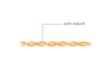
What structural and functional differences exist between cilia and flagella? |
In eukaryotes, both cilia and flagella have a 9+2 method of organization. This consists of 9 microtubule pairs arranged in a circle surrounding 2 single microtubules. |
Describe the composition of the plasma membrane. | It consists of a phospholipid bilayer, with polar heads on the exterior (pointing toward the extracellular fluid and cytoplasm) and nonpolar tails on the interior. The membrane also contains cholesterol, associated large proteins, and sphingolipids, among other components. |
What will happen to a human cell when it is placed in a hypertonic solution? | Water will exit the cell, causing it to shrivel. A hypertonic solution is one that contains a comparatively high amount of solute (salt). Due to osmosis, water will flow from a low-solute to a high-solute environment in an attempt to equalize the solute concentrations. |
What is a concentration gradient, and what is its biological significance? | It is a difference in the amount of solute molecules per unit volume between one region and another. Molecules are prone to moving down their concentration gradient. For example, if the outside of a cell contains a much higher concentration of glucose than the inside, glucose will tend to move into the cell (if possible). If the membrane is impermeable to glucose, water will tend to move out instead. |
If a normal human cell is placed in a container of pure water, what will result? | The cell will swell, possibly to the point of lysing (rupturing). Since pure water contains no solute, it is hypotonic in comparison to the cell. Water will travel down its concentration gradient from the exterior of the cell to the interior. |
What is facilitated diffusion, and is it an active or passive process? |
|
Define: endocytosis | It is a form of transport in which a material is engulfed by the cell membrane, then enters the cell in a vesicle. Endocytosis requires the expenditure of energy. Phagocytosis and pinocytosis are both subtypes of endocytosis. |
Define: exocytosis | It is a form of transport in which a material is packaged into a vesicle which then fuses with the cell membrane. This process, which requires energy, allows the material to be exported from the cell. Waste products and secreted hormones often leave a cell via exocytosis. |
Define: pinocytosis | It is a type of endocytosis. In this process, the cell membrane engulfs the extracellular fluid, as well as the small particles it contains, in a vesicle. Other types of endocytosis include phagocytosis and receptor-mediated endocytosis. |
What features characterize tight junctions? | They are composed of multiple proteins, including claudins, and form an impermeable seal between adjacent cells. This prevents fluid and solutes from going "around" the cell to enter a cavity. Tight junctions are found between cells in the same epithelial layer. |
What features characterize desmosomes? | They are composed of intermediate filaments and are found at localized regions throughout a cell's membrane. They generally attach one epithelial or cell layer to another. Desmosomes are found in epithelial and muscle tissue. |
What features characterize gap junctions? | They are small channels formed from connexin proteins. They allow small solutes and fluid to pass from one cell to another. While gap junctions are found between most cells, they are generally associated with cells that must communicate or function together, like neurons. |
How do eukaryotic and prokaryotic cells differ with respect to organelles? | Unlike eukaryotes, prokaryotes lack a nucleus, as well as all membrane-bound organelles. Note that membrane-bound organelles include mitochondria, lysosomes, the ER, and the Golgi apparatus, but not ribosomes. Prokaryotes do contain ribosomes, a fact that may appear on the AP Biology exam. |
How do eukaryotic and prokaryotic organisms differ in their cellular organization? | Prokaryotes are always unicellular, while eukaryotes can be either unicellular or multicellular. One common example of a unicellular eukaryote is yeast, a fungus. Most other single-celled eukaryotes are classified as protists. |
Determine if an organism with the following traits is a prokaryote or a eukaryote:
| eukaryote Only a eukaryote would possess mitochondria, since prokaryotes lack membrane-bound organelles. Eukaryotes also have linear, not circular, chromosomes. Note that both eukaryotes and prokaryotes can be unicellular. |
In animal cells, which organelle serves as the location for DNA in the form of linear chromosomes? | The nucleus. It is also the site of DNA replication and transcription. While the mitochondria also include DNA, mitochondrial DNA is found in small circular chromosomes, not linear ones. |
Which organelle has two subunits and serves as the location for protein synthesis? | The ribosome. Ribosomes are small organelles found in both eukaryotic and prokaryotic cells. At these organelles, proteins are synthesized (translated). A typical ribosome includes a small and a large subunit, although the sizes of these subunits vary depending on the type of cell. |