2024 WGU D236 Pathophysiology Practice Exam With Answers (134 Solved Questions)

2024 WGU D236 Pathophysiology Practice Exam With Answers is an excellent resource for reviewing and analyzing past exam trends.

Ella Hall
Contributor
4.4
47
5 months ago
Preview (16 of 50 Pages)
100%
Purchase to unlock

Page 1

2024 WGU D236 Pathophysiology Practice Exam With Answers (134 Solved Questions) - Page 1 preview image

Loading page image...

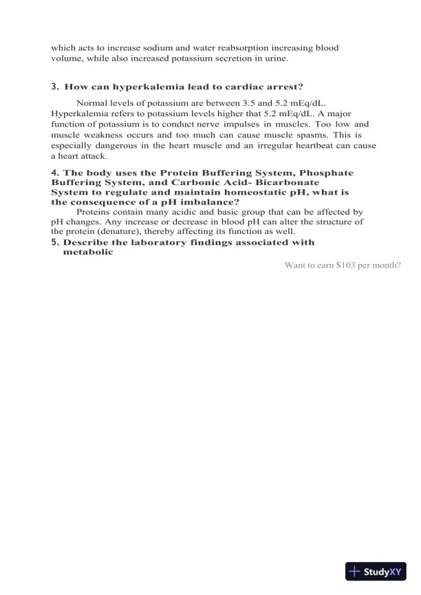
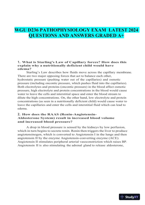
WGUD236PATHOPHYSIOLOGY EXAMLATEST 2024QUESTIONS AND ANSWERSGRADED A+1.What is Starling's Law of Capillary forces? How does thisexplain why a nutritionally deficient child would haveedema?Starling’sLaw describes how fluids move across the capillary membrane.There are two major opposing forces that act to balance each other,hydrostatic pressure (pushing water out of the capillaries) and osmoticpressure (including oncontic pressure, which pushes fluid into the capillaries).Both electrolytes and proteins (oncontic pressure) in the blood affect osmoticpressure, high electrolyte and protein concentrations in the blood would causewater to leave the cells and interstitial space and enter the blood stream todilute the high concentrations. On, the other hand, low electrolyte and proteinconcentrations (as seen in a nutritionally deficient child) would cause water toleave the capillaries and enter the cells and interstitial fluid which can lead toedema.2.How does the RAAS (Renin-Angiotensin-Aldosterone System) result in increased blood volumeand increased blood pressure?A drop in blood pressure is sensed by the kidneys by low perfusion,which in turn begins to secrete renin. Renin then triggers the liver to produceangiotensinogen, which is converted to Angiotensin I in the lungs and thenangiotensin II by the enzyme Angiotensin-converting enzyme (ACE).Angiotensin II stimulates peripheral arterial vasoconstriction which raises BP.Angiotensin II is also stimulating the adrenal gland to release aldosterone,

Page 2

2024 WGU D236 Pathophysiology Practice Exam With Answers (134 Solved Questions) - Page 2 preview image

Loading page image...

Page 3

2024 WGU D236 Pathophysiology Practice Exam With Answers (134 Solved Questions) - Page 3 preview image

Loading page image...

which acts to increase sodium and water reabsorption increasing bloodvolume, while also increased potassium secretion in urine.3.How can hyperkalemia lead to cardiac arrest?Normal levels of potassium are between 3.5 and 5.2 mEq/dL.Hyperkalemia refers to potassium levels higher that 5.2 mEq/dL. A majorfunction of potassium is to conduct nerve impulses in muscles. Too low andmuscle weakness occurs and too much can cause muscle spasms. This isespecially dangerous in the heart muscle and an irregular heartbeat can causea heart attack.4.The body uses the Protein Buffering System, PhosphateBuffering System, and Carbonic Acid- BicarbonateSystem to regulate and maintain homeostatic pH, what isthe consequence of a pH imbalance?Proteins contain many acidic and basic group that can be affected bypH changes. Any increase or decrease in blood pH can alter the structure ofthe protein (denature), thereby affecting its function as well.5.Describe the laboratory findings associated withmetabolicWant to earn $103 per month?

Page 4

2024 WGU D236 Pathophysiology Practice Exam With Answers (134 Solved Questions) - Page 4 preview image

Loading page image...

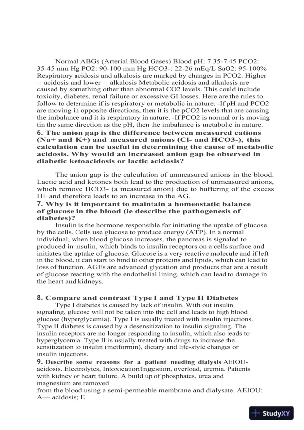
acidosis,metabolicalkalosis,respiratoryacidosisandrespiratory alkalosis. (ie relative pH and CO2 levels).

Page 5

2024 WGU D236 Pathophysiology Practice Exam With Answers (134 Solved Questions) - Page 5 preview image

Loading page image...

Normal ABGs (Arterial Blood Gases) Blood pH: 7.35-7.45 PCO2:35-45 mm Hg PO2: 90-100 mm Hg HCO3-: 22-26 mEq/L SaO2: 95-100%Respiratory acidosis and alkalosis are marked by changes in PCO2. Higher=acidosis and lower=alkalosis Metabolic acidosis and alkalosis arecaused by something other than abnormal CO2 levels. This could includetoxicity, diabetes, renal failure or excessive GI losses. Here are the rules tofollow to determine if is respiratory or metabolic in nature. -If pH and PCO2are moving in opposite directions, then it is the pCO2 levels that are causingthe imbalance and it is respiratory in nature. -If PCO2 is normal or is movingtin the same direction as the pH, then the imbalance is metabolic in nature.6.The anion gap is the difference between measured cations(Na+ and K+) and measured anions (Cl- and HCO3-), thiscalculation can be useful in determining the cause of metabolicacidosis. Why would an increased anion gap be observed indiabetic ketoacidosis or lactic acidosis?The anion gap is the calculation of unmeasured anions in the blood.Lactic acid and ketones both lead to the production of unmeasured anions,which remove HCO3- (a measured anion) due to buffering of the excessH+ and therefore leads to an increase in the AG.7.Why is it important to maintain a homeostatic balanceof glucose in the blood (ie describe the pathogenesis ofdiabetes)?Insulin is the hormone responsible for initiating the uptake of glucoseby the cells. Cells use glucose to produce energy (ATP). In a normalindividual, when blood glucose increases, the pancreas is signaled toproduced in insulin, which binds to insulin receptors on a cells surface andinitiates the uptake of glucose. Glucose is a very reactive molecule and if leftin the blood, it can start to bind to other proteins and lipids, which can lead toloss of function. AGEs are advanced glycation end products that are a resultof glucose reacting with the endothelial lining, which can lead to damage inthe heart and kidneys.8.Compare and contrast Type I and Type II DiabetesType I diabetes is caused by lack of insulin. With out insulinsignaling, glucose will not be taken into the cell and leads to high bloodglucose (hyperglycemia). Type I is usually treated with insulin injections.Type II diabetes is caused by a desensitization to insulin signaling. Theinsulin receptors are no longer responding to insulin, which also leads tohyperglycemia. Type II is usually treated with drugs to increase thesensitization to insulin (metformin), dietary and life-style changes orinsulin injections.9.Describe some reasons for a patient needing dialysisAEIOU-acidosis. Electrolytes, Intoxication/Ingestion,overload, uremia. Patientswith kidney or heart failure. A build up of phosphates, urea andmagnesium are removedfrom the blood using a semi-permeable membrane and dialysate. AEIOU:Aacidosis; E

Page 6

2024 WGU D236 Pathophysiology Practice Exam With Answers (134 Solved Questions) - Page 6 preview image

Loading page image...

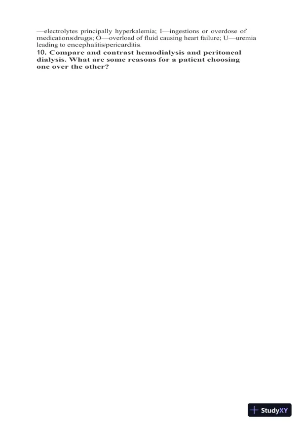
electrolytes principally hyperkalemia; Iingestions or overdose ofmedications/drugs;Ooverload of fluid causing heart failure; Uuremialeading toencephalitis/pericarditis.10.Compare and contrast hemodialysis and peritonealdialysis. What are some reasons for a patient choosingone over the other?

Page 7

2024 WGU D236 Pathophysiology Practice Exam With Answers (134 Solved Questions) - Page 7 preview image

Loading page image...

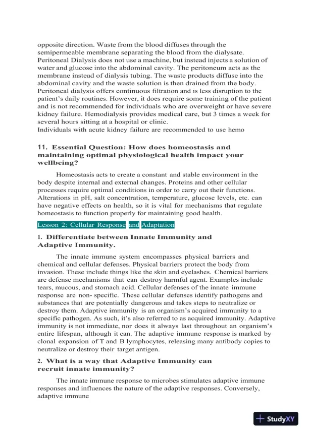
Hemodialysis uses a machine to pump blood from the body in onetube while dialysate (made of water, electrolytes and salts) is pumped in theseparate tube in the

Page 8

2024 WGU D236 Pathophysiology Practice Exam With Answers (134 Solved Questions) - Page 8 preview image

Loading page image...

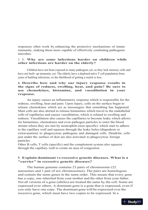
opposite direction. Waste from the blood diffuses through thesemipermeable membrane separating the blood from the dialysate.Peritoneal Dialysis does not use a machine, but instead injects a solution ofwater and glucose into the abdominal cavity. The peritoneum acts as themembrane instead of dialysis tubing. The waste products diffuse into theabdominal cavity and the waste solution is then drained from the body.Peritoneal dialysis offers continuousfiltration and is less disruption to thepatient’s daily routines. However, it doesrequire some training of the patientand is not recommended for individuals who are overweight or have severekidney failure. Hemodialysis provides medical care, but 3 times a week forseveral hours sitting at a hospital or clinic.Individuals with acute kidney failure are recommended to use hemo11.Essential Question: How does homeostasis andmaintaining optimal physiological health impact yourwellbeing?Homeostasis acts to create a constant and stable environment in thebody despite internal and external changes. Proteins and other cellularprocesses require optimal conditions in order to carry out their functions.Alterations in pH, salt concentration, temperature, glucose levels, etc. canhave negative effects on health, so it is vital for mechanisms that regulatehomeostasis to function properly for maintaining good health.Lesson2:CellularResponseandAdaptation1.Differentiate between Innate Immunity andAdaptive Immunity.The innate immune system encompasses physical barriers andchemical and cellular defenses. Physical barriers protect the body frominvasion. These include things like the skin and eyelashes. Chemical barriersare defense mechanisms that can destroy harmful agent. Examples includetears, mucous, and stomach acid. Cellular defenses of the innate immuneresponse are non- specific. These cellular defenses identify pathogens andsubstances that are potentially dangerous and takes steps to neutralize ordestroy them. Adaptive immunityis an organism’s acquired immunity to aspecific pathogen. As such, it’s also referred to as acquired immunity. Adaptiveimmunity is not immediate, nor does it always last throughout anorganism’sentire lifespan, although it can. The adaptive immune response is marked byclonal expansion of T and B lymphocytes, releasing many antibody copies toneutralize or destroy their target antigen.2.What is a way that Adaptive Immunity canrecruit innate immunity?The innate immune response to microbes stimulates adaptive immuneresponses and influences the nature of the adaptive responses. Conversely,adaptive immune

Page 9

2024 WGU D236 Pathophysiology Practice Exam With Answers (134 Solved Questions) - Page 9 preview image

Loading page image...

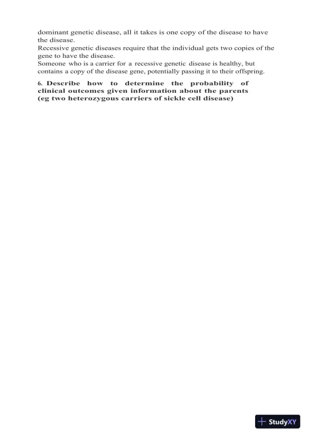
responses often work by enhancing the protective mechanisms of innateimmunity, making them more capable of effectively combating pathogenicmicrobes.3.3.Why are some infections harder on children whileother infections are harder on the elderly?Children have not been exposed to many pathogens yet, so they lack memory cells andhave not built- up immunity yet. The elderly have a depleted naïve T cell population fromyears of battling infections, so the likelihood of getting a match is less.4.Describe how and why our injury response results inthe signs of redness, swelling, heat, and pain? Be sure tousechemokines,histamine,andvasodilationinyourresponse.An injury causes an inflammatory response which is responsible for theredness, swelling, heat and pain. Upon injury, cells on the surface begin torelease chemokines which act as messengers that something has happened.Mast cells are also alerted to release histamines which travel to the endothelialcells of capillaries and causes vasodilation, which is related to swelling andredness. Vasodilation also causes the capillaries to become leaky which allowsfor histamines, chemokines and even pathogen particles to enter the bloodstream where they are met by neutrophils (non-specific) which start to adhereto the capillary wall and squeeze through the leaky holes (diapedesis orextravasation) to phagocytose pathogens and damaged cells. Dendritic cellsjust under the surface of skin are also activated to phagocytose foreignparticles.Other B cells, T cells (specific) and the complement system also squeezethrough the capillary wall to create an area of congestion.5.Explain dominant vs recessive genetic diseases. What is a"carrier" in recessive genetic diseases?The human genome contains 23 pairs of chromosome (22autosomes and 1 pair of sex chromosomes). The pairs are homologousand contain the same genes in the same order. This means that every genehas a copy, oneinherited from your mother and the other from your father.Not all versions ofa gene (alleles) are treated the same by the cell. Some areexpressed over others. A dominant gene is a gene that is expressed, even ifyou only have one copy. The dominant gene will be expressed over therecessive gene, which must have two copies to be expressed. In a

Page 10

2024 WGU D236 Pathophysiology Practice Exam With Answers (134 Solved Questions) - Page 10 preview image

Loading page image...

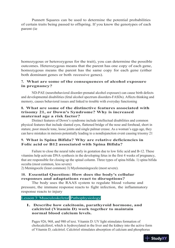
dominant genetic disease, all it takes is one copy of the disease to havethe disease.Recessive genetic diseases require that the individual gets two copies of thegene to have the disease.Someone who is a carrier for a recessive genetic disease is healthy, butcontains a copy of the disease gene, potentially passing it to their offspring.6.Describehowtodeterminetheprobabilityofclinical outcomes given information about the parents(eg two heterozygous carriers of sickle cell disease)

Page 11

2024 WGU D236 Pathophysiology Practice Exam With Answers (134 Solved Questions) - Page 11 preview image

Loading page image...

Punnett Squares can be used to determine the potential probabilitiesof certain traits being passed to offspring. If you know the genotypes of eachparent (iehomozygous or heterozygous for the trait), you can determine the possibleoutcomes. Heterozygous means that the parent has one copy of each gene,homozygous means the parent has the same copy for each gene (eitherboth dominant genes or both recessive genes).7.What are some of the consequences of alcohol exposurein pregnancy?ND-PAE (neurobehavioral disorder-prenatal alcohol exposure) can cause birth defectsand developmental disabilities (fetal alcohol spectrum disorders-FASDs). Affects thinking andmemory, causes behavioral issues and linked to trouble with everyday functioning8.What are some of the distinctive features associated withtrisomy 21, or Down's Syndrome? Why is increasedmaternal age a risk factor?Distinct features ofDown’ssyndrome include intellectual disabilities and commonphysical features that include slanted eyes, flattened bridge of the nose and forehead, short instature, poor muscle tone, loose joints and single palmar crease. As awoman’seggs age, theycan have mistakes in meiosis potentially leading to a nondisjunction event causing trisomy 219.What is Spina Bifida? Why are relative deficiencies inFolic acid or B12 associated with Spina Bifida?Failure to close the neural tube early in gestation due to low folic acid and B-12. Thesevitamins help activate DNA synthesis in the developing fetus in the first 4 weeks of pregnancy,that are responsible for closing up the spinal column. Three types of spina bifida. 1) spina bifidaocculta (most common, less severe)2) Meningocele (least common) 3) Myelomeningocele (most severe).10.Essential Question: How does the body's cellularresponses and adaptations react to disruptions?The body uses the RAAS system to regulate blood volume andpressure, the immune response reacts to fight infection, the inflammatoryresponse reacts to injuryLesson3:MusculoskeletalPathophysiology1.Describe how calcitonin, parathyroid hormone, andcalcitriol (Vitamin D) work together to maintainnormal blood calcium levels.Pages 926, 968, and 980 of text. Vitamin D: UV light stimulates formation ofcholecalciferol, which is hydroxylated in the liver and the kidney into the active formof Vitamin D, calcitriol. Calcitriol stimulates absorption of calcium and phosphorus

Page 12

2024 WGU D236 Pathophysiology Practice Exam With Answers (134 Solved Questions) - Page 12 preview image

Loading page image...

from the GI tract in the intestine and phosphate in the kidney. Calcitriol increases thecalcification of osteoid. Calcitriol also stimulates the formation of bone by raising thelevels of calcium and phosphorus in the blood. Low vitamin D levels can causehypocalcemia, which stimulates the parathyroid gland to release parathyroid hormone(PTH). PTH stimulates osteoclasts to resorb bone calcium to increase blood calciumlevels. PTH also stimulates

Page 13

2024 WGU D236 Pathophysiology Practice Exam With Answers (134 Solved Questions) - Page 13 preview image

Loading page image...

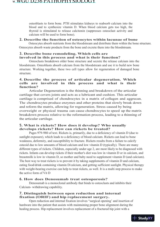
WWGU D236 PATHOPHYSIOLOGYosteoblasts to form bone. PTH stimulates kidneys to reabsorb calcium into theblood and to synthesize vitamin D. When blood calcium gets too high, thethyroid is stimulated to release calcitonin (suppresses osteoclast activity andcalcium will be used to form bone).2.Describe the function of osteocytes within lacunae of boneOsteocytes absorb nutrients from the bloodstream and distribute them within the bone structure.Osteocytes absorb waste products from the bone and excrete them into the bloodstream.3.Describe bone remodeling. Which cells areinvolved in this process and what is their function?Osteoclasts breakdown older bone structure and secrete the release calcium into thebloodstream. Osteoblasts absorb calcium from the bloodstream and use it to build new bonestructure. Working together, these two cell types allow for regeneration of damaged bonestructure.4.Describe the process of articular degeneration. Whichcellsareinvolvedinthisprocessandwhatistheirfunction?Articular Degeneration is the thinning and breakdown of the articularcartilage that covers joints and acts as a lubricant and cushion. This articularcartilage is comprised of chondrocytes in a matrix of collagen and aggrecan.The chondrocytes produce enzymes and other proteins that slowly break downand reform the matrix, allowing for regeneration. Stress caused by beingoverweight or physical trauma can cause chondrocytes to speed up the matrixbreakdown process relative to the reformation process, leading to a thinning ofthe articular cartilage.5.What is rickets? How does it develop? Who usuallydevelops rickets? How can rickets be treated?Pages 979-980 of text. Rickets is, primarily, due to a deficiency of vitamin D (due tosunlight exposure), which leads to a deficiency of blood calcium. Rickets can lead to boneweakness, deformity, and susceptibility to fracture. Rickets results from a failure to calcifyosteoid due to low amounts of blood calcium and low vitamin D (typically). There are manydifferent types of rickets. Children, especially under age 2, are most likely to be diagnosed withrickets. Infants can develop rickets if their mother's diet was low in vitamin D or in calcium, andbreastmilk is low in vitamin D, so mother and baby need to supplement vitamin D (and calcium).The best way to treat rickets is to prevent it by taking supplements of vitamin D and calcium,eating food/drink containing vitamin D/calcium, and getting sufficient sunlight. Physical therapywith weight-bearing exercise can help to treat rickets, as well. It is a multi step process to makethe active form of Vit D6.How does Denosumab treat osteoporosis?Denosumab is a monoclonal antibody that binds to osteoclasts and inhibits theirCalcium- withdrawing capability.7.Distinguish between open reduction and internalfixation (ORIF) and hip replacement surgery.Open reduction and internal fixation involves―surgicalopening’and insertion ofhardware into the patient that assists with maintaining proper bone alignment during thehealing process. Hip replacement involves replacement of a fractured hip joint with a

Page 14

2024 WGU D236 Pathophysiology Practice Exam With Answers (134 Solved Questions) - Page 14 preview image

Loading page image...

prosthesis.8.What is degenerative disc disease? What are some of theanatomical features of degenerative disc disease? Whatare some symptoms of

Page 15

2024 WGU D236 Pathophysiology Practice Exam With Answers (134 Solved Questions) - Page 15 preview image

Loading page image...

lumbar vs. cervical degenerative disc disease??

Page 16

2024 WGU D236 Pathophysiology Practice Exam With Answers (134 Solved Questions) - Page 16 preview image

Loading page image...

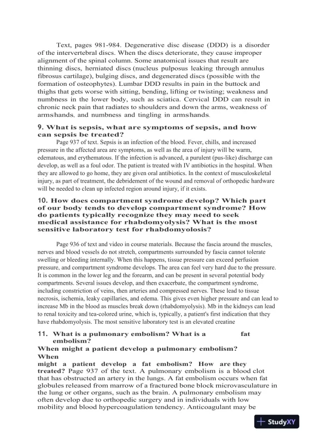
Text, pages 981-984. Degenerative disc disease (DDD) is a disorderof the intervertebral discs. When the discs deteriorate, they cause improperalignment of the spinal column. Some anatomical issues that result arethinning discs, herniated discs (nucleus pulposus leaking through annulusfibrosus cartilage), bulging discs, and degenerated discs (possible with theformation of osteophytes). Lumbar DDD results in pain in the buttock andthighs that gets worse with sitting, bending, lifting or twisting; weakness andnumbness in the lower body, such as sciatica. Cervical DDD can result inchronic neck pain that radiates to shoulders and down the arms, weakness ofarms/hands,and numbness and tingling inarms/hands.9.What is sepsis, what are symptoms of sepsis, and howcan sepsis be treated?Page 937 of text. Sepsis is an infection of the blood. Fever, chills, and increasedpressure in the affected area are symptoms, as well as the area of injury will be warm,edematous, and erythematous. If the infection is advanced, a purulent (pus-like) discharge candevelop, as well as a foul odor. The patient is treated with IV antibiotics in the hospital. Whenthey are allowed to go home, they are given oral antibiotics. In the context of musculoskeletalinjury, as part of treatment, the debridement of the wound and removal of orthopedic hardwarewill be needed to clean up infected region around injury, if it exists.10.How does compartment syndrome develop? Which partof our body tends to develop compartment syndrome? Howdo patients typically recognize they may need to seekmedical assistance for rhabdomyolysis? What is the mostsensitive laboratory test for rhabdomyolosis?Page 936 of text and video in course materials. Because the fascia around the muscles,nerves and blood vessels do not stretch, compartments surrounded by fascia cannot tolerateswelling or bleeding internally. When this happens, tissue pressure can exceed perfusionpressure, and compartment syndrome develops. The area can feel very hard due to the pressure.It is common in the lower leg and the forearm, and can be present in several potential bodycompartments. Several issues develop, and then exacerbate, the compartment syndrome,including constriction of veins, then arteries and compressed nerves. These lead to tissuenecrosis, ischemia, leaky capillaries, and edema. This gives even higher pressure and can lead toincrease Mb in the blood as muscles break down (rhabdomyolysis). Mb in the kidneys can leadto renal toxicity and tea-colored urine, which is, typically, a patient's first indication that theyhave rhabdomyolysis. The most sensitive laboratory test is an elevated creatine11.What is a pulmonary embolism? What is afatembolism?When might a patient develop a pulmonary embolism?Whenmightapatientdevelopafatembolism?Howare theytreated?Page 937 of the text. A pulmonary embolism is a blood clotthat has obstructed an artery in the lungs. A fat embolism occurs when fatglobules released from marrow of a fractured bone block microvasculature inthe lung or other organs, such as the brain. A pulmonary embolism mayoften develop due to orthopedic surgery and in individuals with lowmobility and blood hypercoagulation tendency. Anticoagulant may be
Preview Mode

This document has 50 pages. Sign in to access the full document!

Study Now!

XY-Copilot AI
Unlimited Access
Secure Payment
Instant Access
24/7 Support
Document Chat

Document Details

Related Documents

View all