Oral Pathology for the Dental Hygienist 7th Edition Test Bank

Gain an edge in your exam with Oral Pathology for the Dental Hygienist 7th Edition Test Bank, covering core subjects with a structured learning approach.

Henry Walker
Contributor
4.3
42
5 months ago
Preview (16 of 228 Pages)
100%
Purchase to unlock

Page 1

Oral Pathology for the Dental Hygienist 7th Edition Test Bank - Page 1 preview image

Loading page image...

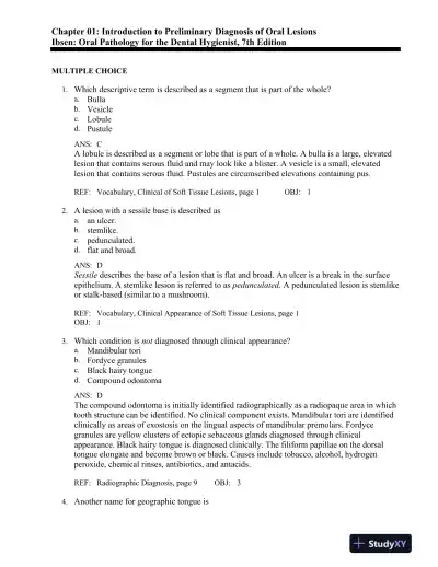
Chapter 01: Introduction to Preliminary Diagnosis of Oral LesionsIbsen: Oral Pathology for the Dental Hygienist, 7th EditionMULTIPLE CHOICE1.Which descriptive term is described as a segment that is part of the whole?a.Bullab.Vesiclec.Lobuled.PustuleANS:CA lobule is described as a segment or lobe that is part of a whole. A bulla is alarge, elevatedlesion that contains serous fluid and may look like a blister. A vesicle is a small, elevatedlesion that contains serous fluid. Pustules are circumscribed elevations containing pus.REF:Vocabulary, Clinical of Soft Tissue Lesions, page 1OBJ:12.A lesion with a sessile base is described asa.an ulcer.b.stemlike.c.pedunculated.d.flat and broad.ANS:DSessiledescribes the base of a lesion that is flat and broad. An ulcer is a break in the surfaceepithelium. A stemlike lesion is referred to aspedunculated. A pedunculated lesion is stemlikeor stalk-based (similar to a mushroom).REF:Vocabulary, Clinical Appearance of Soft Tissue Lesions, page 1OBJ:13.Which condition isnotdiagnosed through clinical appearance?a.Mandibular torib.Fordyce granulesc.Black hairy tongued.Compound odontomaANS:DThe compound odontoma is initially identified radiographically as a radiopaque area in whichtooth structure can be identified. No clinical component exists. Mandibular tori are identifiedclinically as areas of exostosis on the lingual aspects of mandibular premolars. Fordycegranules are yellow clusters of ectopic sebaceous glands diagnosed through clinicalappearance. Black hairy tongue is diagnosed clinically. The filiform papillae on the dorsaltongue elongate and become brown or black. Causes include tobacco, alcohol, hydrogenperoxide, chemical rinses, antibiotics, and antacids.REF:Radiographic Diagnosis, page 9OBJ:34.Another name for geographic tongue is

Page 2

Oral Pathology for the Dental Hygienist 7th Edition Test Bank - Page 2 preview image

Loading page image...

Page 3

Oral Pathology for the Dental Hygienist 7th Edition Test Bank - Page 3 preview image

Loading page image...

a.median rhomboid glossitis.b.benign migratory glossitis.c.fissured tongue.d.black hairy tongue.ANS:BBenign migratory glossitis is another name for geographic tongue. Research suggests thatmedian rhomboid glossitis is associated with a chronic fungal infection fromCandidaalbicans. Sometimes the condition resolves with antifungal therapy. Fissured tongue is seen in5% of the population. It is a variant of normal. Genetic factors are typically associated withthe condition. Black hairy tongue is caused by a reaction to chemicals, tobacco, hydrogenperoxide, or antacids. The filiform papillae on the dorsal tongue become elongated and aredark brown to black.REF:Geographic Tongue, page 24OBJ:75.This bony hard structure in the midline of the hard palate is genetic in origin and inherited inan autosomal dominant manner. The diagnosis is made through clinical appearance. Whichcondition is suspected?a.Palatal cystb.Torus palatinusc.Mixed tumord.RanulaANS:BA torus palatinus is developmental and bony hard and is found on the midline of the palate.Diagnosis is made on the basis of clinical appearance. A palatal cyst appears radiolucent on aradiographic examination and is not diagnosed through clinical appearance. A mixed tumor orpleomorphic adenoma is a benign tumor of salivary gland origin, found unilaterally off themidline of the hard palate. It is composed of tumor tissue that is not bony hard to palpation.Ranulais a term used for a mucocele-like lesion that forms unilaterally on the floor of themouth.REF:Torus Palatinus, page 21OBJ:46.The gray-white opalescent film seen on the buccal mucosa of 85% of black adults is a variantof normal that requires no treatment and is termeda.linea alba.b.leukoedema.c.leukoplakia.d.white sponge nevus.ANS:BLeukoedema is a diffuse opalescence most commonly seen on the buccal mucosa in blackindividuals. Linea alba is a “white line” that extends anteroposteriorly on the buccal mucosaalong the occlusal plane. It is most prominent in patients who have a clenching or grindinghabit.Leukoplakiais a clinical term for a white lesion, the cause of which is unknown. Whitesponge nevus is a genetic (autosomal dominant) trait. Clinically, it is characterized by a softwhite, folded (orcorrugated) oral mucosa. A thick layer of keratin produces the whitening.REF:Leukoedema, page 23OBJ:8

Page 4

Oral Pathology for the Dental Hygienist 7th Edition Test Bank - Page 4 preview image

Loading page image...

7.Which condition most likely responds to therapeutic diagnosis?a.Angular cheilitisb.Amelogenesis imperfectac.Paget diseased.Stafne bone cystANS:AAngular cheilitis most commonly responds to antifungal therapy once nutritional deficiencieshave been ruled out. Amelogenesis imperfecta is a genetic condition associated with abnormaldevelopment of the enamel. Paget disease is a chronic metabolic bone disease. A highlyelevated serum alkaline phosphatase level contributes significantly to the diagnosis. A Stafnebone cyst is determined through surgical diagnosis in which entrapped salivary gland tissue isidentified.REF:Therapeutic Diagnosis, page 18OBJ:38.The gingival enlargement in this patient was caused by a calcium channel blocker. Whichmedication is the likely cause?a.Dilantinb.Nifedipinec.Quinidined.ClozapineANS:BNifedipine is a calcium channel blocker. Dilantin is an anticonvulsant used to prevent orcontrol seizures. Quinidine is an antiarrhythmic agent used to treat cardiac arrhythmias.Clozapine is an antipsychotic used in the management of psychotic symptoms inschizophrenia.REF:Historical Diagnosis, Fig. 1.38, page 17OBJ:39.Radiographic features, including cotton-wool radiopacities and hypercementosis, areespecially helpful in the diagnosis ofa.Paget disease.b.dentinogenesis imperfecta.c.anemia.d.diabetes.ANS:APaget disease is a chronic metabolic bone disease. Radiographically, cotton-woolradiopacitiesand hypercementosis are characteristic features. Dentinogenesis imperfecta is a geneticcondition involving a defect in the development of dentin. Anemia, a decrease in red bloodcells, requires blood tests to determine the etiologic factors. Diabetes is a chronic disorder ofcarbohydrate metabolism characterized by abnormally high blood glucose levels.REF:Laboratory Diagnosis, Fig. 1.40, pages 16, 18OBJ:310.In internal resorption, the radiolucency seen on radiographic examination is usuallya.well circumscribed.b.diffuse.

Page 5

Oral Pathology for the Dental Hygienist 7th Edition Test Bank - Page 5 preview image

Loading page image...

c.multilocular.d.unilocular.ANS:BDiffuse borders are ill defined, making it impossible to detect the exact parameters of thelesion. Therefore treatment is more difficult.Well circumscribeddescribes borders that arespecifically defined. Exact margins of the lesion are identified.Multilocularhas also beendescribed as resembling “soap bubbles”; lobes seem to fuse together to make up the lesion.This term has been used to describe the odontogenic keratocyst.Unilocularmeans having onecompartment or unit that is well defined. This term is often used to describe the radicular cyst.REF:Vocabulary, Radiographic Terms Used to Describe Lesions in Bone, page 5OBJ:111.Which condition is diagnosed through clinical appearance?a.Fordyce granulesb.Unerupted mesiodensc.Periapical cemento-osseous dysplasiad.Traumatic bone cystANS:AFordyce granules are diagnosed on the basis of their clinical appearance. They are ectopicsebaceous glands seen on the lips and buccal mucosa. Clinically, they appear as yellowlobules in clusters and are considered a variant of normal. Unerupted mesiodens requires aradiographic image for diagnosis. Periapical cemento-osseous dysplasia requires aradiographic image, specific patient history, and a pulp test to evaluate tooth vitality.Traumatic bone cyst requires a radiographic image and surgical intervention to establish adiagnosis.REF:Clinical Diagnosis, page 7 | Fordyce Granules, page 20OBJ:312.Retrocuspid papillae are located on thea.palate.b.floor of the mouth.c.gingival margin of the lingual aspect of mandibular cuspids.d.canine eminence.ANS:CRetrocuspid papillae are located on the gingival margin of the lingual aspect of mandibularcuspids. Retrocuspid papillae are not located on the palate. Retrocuspid papillae are notlocated on the floor of the mouth. Retrocuspid papillae are not located on the canineeminence.REF:Retrocuspid Papilla, page 22OBJ:313.Which condition isnotconsidered a variant of normal?a.Fordyce granulesb.Leukoedemac.Linea albad.Pyogenic granulomaANS:D

Page 6

Oral Pathology for the Dental Hygienist 7th Edition Test Bank - Page 6 preview image

Loading page image...

Pyogenic granuloma is a reactive inflammatory response to injury. It is not a variant ofnormal. Fordyce granules are seen in more than 80% of adults over 20 years of age and areconsidered a variant of normal. Leukoedema is observed in about 85% of black individualsand is considered a variant of normal. Linea alba is located on the buccal mucosa along theocclusal plane of the teeth. It is most prominent in patients who have a clenching or bruxismhabit. It is so common that it is considered a variant of normal.REF:Box 1-1, Case Study, page 20 | Variants of Normal, pages 21-23OBJ:614.When antifungal therapy is used to treat angular cheilitis, which diagnostic process is beingapplied?a.Microscopicb.Laboratoryc.Surgicald.TherapeuticANS:DTherapeutic diagnosis is used here in the treatment and management of angular cheilitis,which is most commonly a fungal condition. A careful patient history should be obtained torule out a contributory nutritional deficiency. Microscopic diagnosis requires a biopsy.Laboratory diagnosis involves the use of clinical laboratory tests, including blood chemistriesand urinalysis. Surgical diagnosis requires surgical intervention.REF:Therapeutic Diagnosis, page 18OBJ:315.A lesion with a stemlike base is described asa.sessile.b.macular.c.pedunculated.d.lobulated.ANS:CPedunculatedmeans the lesion has a stemlike or stalklike base similar to that of a mushroom.A sessile base is broad and flat. A macular lesion is flat, does not protrude, and isdistinguished by its color. A freckle is an example of a macule.Lobulatedmeans the lesionconsists of lobules making up the whole.REF:Vocabulary, Clinical Appearance of Soft Tissue Lesions, page 1OBJ:816.A small, circumscribed lesion, usually less than 1 cm in diameter, that is elevated or protrudesabove the surface of normal surrounding tissue is termed aa.papule.b.macule.c.vesicle.d.bulla.ANS:A

Page 7

Oral Pathology for the Dental Hygienist 7th Edition Test Bank - Page 7 preview image

Loading page image...

A papule is defined as a small circumscribed lesion, usually less than 1 cm in diameter, that iselevated or protrudes above the surface of normal surrounding tissue. A macule is flat anddoes not protrude above the surface of normal tissue. A vesicle is elevated and contains serousfluid. A bulla is elevated, contains serous fluid, and looks like a blister.REF:Vocabulary, Clinical Appearance of Soft Tissue Lesions, page 1OBJ:117.Which condition is considered a genetic/inherited disorder?a.Linea albab.Amelogenesis imperfectac.Necrotizing ulcerative gingivitisd.Internal resorptionANS:BAmelogenesis imperfecta represents a group of inherited conditions affecting the enamel ofteeth. Linea alba is a variant of normal characterized by the white line that extendsanteroposteriorly on the buccal mucosa, along the occlusal plane. Necrotizing ulcerativegingivitis is an ulcerating gingival condition caused by anaerobic bacteria. Internal resorptionis usually associated with an inflammatory response in the pulp.REF:Historical Diagnosis, page 15OBJ:318.Ectopic geographic tongue can be found in which location?a.On the lateral border of the tongueb.Within bonec.On mucosal surfaces other than the tongued.The dorsal surface of the tongueANS:CEctopic geographic tongue is also calledstomatitis areata migransand is found on mucosalsurfaces other than the tongue. Geographic tongue may be seen on any surface of the tongue,including the lateral borders. Ectopic geographic tongue is a mucosal condition and isunrelated to the bone. Geographic tongue may be seen on any mucosal surface and iscommonly seen on the dorsal surface of the tongue.REF:Geographic Tongue, page 24OBJ:719.A Stafne bone cyst containsa.salivary gland tissue.b.an empty void.c.inflammatory cells.d.an epithelium-lined cyst containing serous fluid.ANS:AStafne bone cyst is a developmental invagination in the lingual aspect of the mandible that isfilled with salivary gland tissue. An empty void describes the contents of a traumatic bonecyst. A Stafne bone cyst has no inflammatory response. It is developmental and requires notreatment. A Stafne bone cyst is not a true cyst and does not have an epithelial lining. Itcontains normal salivary gland tissue.REF:Surgical Diagnosis, page 18OBJ:3

Page 8

Oral Pathology for the Dental Hygienist 7th Edition Test Bank - Page 8 preview image

Loading page image...

20.Which papillae are elongated in the condition black hairy tongue?a.Circumvallateb.Foliatec.Filiformd.FungiformANS:CThe filiform papillae are composed primarily of keratin and are the papillae that elongate inblack hairy tongue. Circumvallate papillae on the posterior dorsal tongue do not elongate.Foliate papillae are located on the posterior lateral borders of the tongue. They are vertical,slightly exophytic folds of tissue located in the same area as the lingual lymphoid tissue(lingual tonsils) and do not elongate. Fungiform papillae on the dorsal areas do not elongate.REF:Hairy Tongue, page 24OBJ:321.Diagnosis of anemia is best accomplished through which examination?a.Laboratory testsb.Evaluation of the color of the gingivac.Bleeding on probingd.Patient medical historyANS:ALaboratory blood tests provide the best information with which to diagnose anemia. Althoughthe color of the gingiva may be a clinical sign, a diagnosis of anemia, or more specifically thetype of anemia, must be made through laboratory testing. Bleeding on probing is not useful inthe diagnosis of anemia. The patient’s medical history may be helpful in the diagnosis ofanemia, but it is only contributory. The final diagnosis comes from appropriate laboratorytests.REF:Laboratory Diagnosis, page 16OBJ:322.A radiolucency that “scallops around the roots of teeth” is often used to describe whichcondition?a.Traumatic bone cystb.Stafne bone cystc.Lingual mandibular bone concavityd.Median palatine cystANS:AScalloping around the rootsis a term often used to describe the radiographic appearance of atraumatic bone cyst. A Stafne bone cyst usually presents as an oval radiolucency anterior tothe angle of the ramus and inferior to the mandibular canal. A lingual mandibular boneconcavity is the same lesion as a Stafne bone cyst. A median palatine cyst is a developmentalcyst that presents as a unilocular radiolucency found in the midline of the hard palate.REF:Vocabulary, Radiographic Terms Used to Describe Lesions in Bone, page 5OBJ:323.When geographic tongue occurs on the dorsal tongue, the erythematous areas are described asa.inflamed.

Page 9

Oral Pathology for the Dental Hygienist 7th Edition Test Bank - Page 9 preview image

Loading page image...

b.depapillated.c.allergic.d.fungal.ANS:BIn geographic tongue, the erythematous areas on the dorsal tongue are devoid of filiformpapillae and therefore are appropriately described as depapillated. The erythematous areasseen on the dorsal tongue in geographic tongue are not caused primarily by inflammation.Geographic tongue is not an allergic reaction. Geographic tongue is not a fungal infection.REF:Geographic Tongue, page 24OBJ:724.It has been suggested thatCandida albicansis associated with which condition?a.Median rhomboid glossitisb.Linea albac.Leukoedemad.Retrocuspid papillaeANS:AResearch has suggested that median rhomboid glossitis may be associated withCandidaalbicans. Linea alba is a variant of normal. Leukoedema is a variant of normal. Retrocuspidpapillae are developmental and found on the gingiva of the lingual aspect of mandibularcanine teeth.REF:Median Rhomboid Glossitis, page 23OBJ:725.Which term is used to describe a radiographic lesion with borders that are specifically defined,revealing the exact margins and extent of the lesion?a.Unilocularb.Well circumscribedc.Diffused.MultilocularANS:BWell circumscribeddefines a lesion with borders that are specifically defined and in whichone can see the exact margins and extent of the lesion.Unilocularmeans having onecompartment or unit that is well defined (as in a radicular cyst).Diffusedescribes a lesionwith borders that are not well defined. Multilocular lesions are described radiographically asresembling “soap bubbles” (i.e., a lesion with many lobes beyond the confines of one distinctarea).REF:Vocabulary, Radiographic Terms Used to Describe Lesions in Bone, page 5OBJ:126.Fordyce granulesa.are ectopic sebaceous glands.b.are seen on the dorsal surface of the tongue.c.require antibiotic treatment.d.are lesions that require biopsy for diagnosis.ANS:A

Page 10

Oral Pathology for the Dental Hygienist 7th Edition Test Bank - Page 10 preview image

Loading page image...

Fordyce granules are ectopic sebaceous glands. Fordyce granules are seen on thebuccal andlabial mucosa, not the dorsal tongue. Fordyce granules require no treatment. Fordyce granulesare diagnosed through clinical appearance. Biopsy is not necessary.REF:Fordyce Granules, page 20OBJ:427.The base of this lesion is correctly described asa.pedunculated.b.lobule.c.bulla.d.sessile.ANS:DThe base of this lesion is sessile, or broad and flat. A pedunculated lesion hasa stemlike basesimilar to that of a mushroom stem. A lobule is a segment or lobe that is part of the whole. Abulla usually contains serous fluid and looks like a blister.REF:Vocabulary, Clinical Appearance of Soft Tissue Lesions, Fig. 1.5, A, pages 1, 3OBJ:128.This torus palatinus is correctly described asa.bullous.b.papular.c.lobulated.d.nodular.ANS:CThis torus palatinus is lobulated (i.e., lobes that are fused together). Bullous lesions containserous fluid and resemble blisters. A papule is a soft tissue lesion that is elevated above thesurface of normal surrounding tissue. A nodule is a palpable solid lesion found in soft tissue.REF:Vocabulary, Clinical Appearance of Soft Tissue Lesions, Fig. 1.1, pages 1, 2OBJ:129.Another term for an amalgam tattoo is aa.melanoma.b.focal argyrosis.c.nevus.d.multiple myeloma.ANS:BFocal argyrosis is a synonym for amalgam tattoo. A melanoma is a malignant tumor. A nevusis a benign overgrowth of melanocytes. Multiple myeloma is a malignant proliferation ofplasma cells.REF:Clinical Diagnosis, page 7OBJ:230.Which finding isnotapparent in leukoedema?a.Intracellular edema in the spinous cellsb.Acanthosis of the epitheliumc.Generalized opalescence of the buccal mucosad.A white diffuse material on the buccal mucosa that can be wiped off

Page 11

Oral Pathology for the Dental Hygienist 7th Edition Test Bank - Page 11 preview image

Loading page image...

ANS:DA white diffuse material that can be wiped off is significant in diagnosing pseudomembranouscandidiasis. Intracellular edema in the spinous cells is found in leukoedema. Acanthosis of theepithelium is found in leukoedema. Generalized opalescence of the buccal mucosa is seen inleukoedema.REF:Leukoedema, page 23OBJ:831.Which term best describes an elevated, 5-mm soft tissue lesion containing serous fluid?a.Maculeb.Vesiclec.Lobuled.PustuleANS:BA vesicle is a small (less than 1 cm in diameter), elevated lesion that containsserous fluid. Amacule is a flat area usually distinguished by a color different from the surrounding tissue. Alobule is described as a segment or lobe that is part of a whole. A pustule is a circumscribedelevation containing pus.REF:Vocabulary, Clinical Appearance of Soft Tissue Lesions, page 1OBJ:132.A lesion with a pedunculated base is best described asa.an ulcer.b.stemlike.c.pallor.d.broad and flat.ANS:BA stemlike lesion is referred to aspedunculated. An ulcer is a break in the surface epithelium.Pallor is a paleness of the skin or mucosal tissues.Broad and flatdescribes the base of asessile lesion.REF:Vocabulary, Clinical Appearance of Soft Tissue Lesions, page 1OBJ:133.The following lesions can be identified radiographicallyexceptone. Which one is theexception?a.Root resorptionb.Fordyce granulesc.Interproximal dental cariesd.Compound odontomaANS:BFordyce granules are yellow clusters of ectopic sebaceous glands that are diagnosed throughclinical appearance. Root resorption is identified radiographically when the apex of the toothappears shortened or blunted. Interproximal dental caries are seen as radiographicradiolucencies. A compound odontoma is initially identified radiographically as a radiopaquearea in which tooth structure can be identified.

Page 12

Oral Pathology for the Dental Hygienist 7th Edition Test Bank - Page 12 preview image

Loading page image...

REF:Clinical Diagnosis, page 7 | Fordyce Granules, page 20OBJ:334.These reddish-purple clusters observed on the ventral surface of the tongue and diagnosedthrough clinical observation are referred to as which variants of normal conditions?a.Palatal cystb.Mandibular toric.Lingual varicositiesd.RanulaANS:CLingual varicosities are prominent lingual veins usually observed on the ventral and lateralsurfaces of the tongue. A palatal cyst is radiolucent and not diagnosed through clinicalappearance. Mandibular tori are outgrowths of hard dense bone and found on the lingualaspect of the mandible.Ranulais a term used to describe a mucocele-like lesion that formsunilaterally on the floor of the mouth.REF:Lingual Varicosities, page 22OBJ:435.An elevated serum alkaline phosphate level, significant in the diagnosis of Paget disease, isdetermined from which diagnostic category?a.Laboratoryb.Therapeuticc.Clinicald.SurgicalANS:ALaboratory tests, including blood chemistries, can provide information that contributes to adiagnosis. Therapeutic testing applies the principle of diagnosis based on clinical andhistorical information with confirmation by the response of the condition to therapy. Clinicaldiagnosis suggests that the strength of the diagnosis comes from the clinical appearance of thelesion. Surgical diagnosis is based on information about a lesion gained during surgicalintervention.REF:Laboratory Diagnosis, page 16OBJ:236.The following conditions most likely respond to therapeutic diagnosisexceptone. Which oneis theexception?a.Angular cheilitisb.Necrotizing ulcerative gingivitisc.Nutritional deficienciesd.Stafne bone cystANS:DStafne bone cyst, in which entrapped salivary gland tissue is identified, is diagnosed throughsurgical examination. Angular cheilitis commonly responds to antifungal therapy oncenutritional deficiencies have been ruled out. Necrotizing ulcerative gingivitis responds tohydrogen peroxide rinses. Nutritional deficiencies are common conditions diagnosed bytherapeutic means.REF:Surgical Diagnosis, page 18OBJ:3

Page 13

Oral Pathology for the Dental Hygienist 7th Edition Test Bank - Page 13 preview image

Loading page image...

37.The variant of normal coloration seen on the mandibular gingival surface is termeda.lingual varicosities.b.leukoedema.c.melanin pigmentation.d.linea alba.ANS:CMelanin pigmentation is most commonly seen in dark-skinned individuals and givescolor tothe oral mucosa and gingiva. Lingual varicosities are prominent lingual veins observed on theventral and lateral surfaces of the tongue. Leukoedema is a generalized opalescence impartedto the buccal mucosa. Linea alba is a white line that extends anteroposteriorly on the buccalmucosa along the occlusal plane of the teeth.REF:Melanin Pigmentation, Fig. 1.50, pages 21-22OBJ:638.A white lesion that cannot be rubbed off and cannot be diagnosed on the basis of clinicalcharacteristics alone is termeda.leukoplakia.b.dentinogenesis imperfecta.c.erythroplakia.d.squamous cell carcinoma.ANS:ALeukoplakiais a clinical term for a white lesion that cannot be rubbed off and cannot bediagnosed on the basis of clinical characteristics alone. Dentinogenesis imperfecta is a geneticcondition involving a defect in the development of dentin.Erythroplakiais a clinical term fora red lesion that cannot be diagnosed on the basis of clinical features alone. Squamous cellcarcinoma is diagnosed by microscopic evaluation and does not refer to all white lesions thatcannot be rubbed off.REF:Microscopic Diagnosis, page 18OBJ:939.The radiographic appearance of a simple radicular cyst is best described asa.coalescence.b.diffuse.c.multilocular.d.unilocular.ANS:DUnilocularmeans having one compartment or unit that is well defined. This term is often usedto describe a radicular cyst.Coalescencerefers to the process by which parts of a whole jointogether, or fuse, to make one.Diffusedescribes a lesion with ill-defined borders, making itimpossible to detect the exact parameters of the lesion. A multilocular lesion has also beendescribed as “soap bubble”like; the lobes appear to fuse together to make up the lesion.REF:Vocabulary, Radiographic Terms Used to Describe Lesions in Bone, Fig. 1.13, pages 5, 6OBJ:140.A pathologic lesion found frequently in 30-year-old black women that requires a radiographicimage and historical data for diagnosis is termeda.verrucous vulgaris.

Page 14

Oral Pathology for the Dental Hygienist 7th Edition Test Bank - Page 14 preview image

Loading page image...

b.necrotizing ulcerative gingivitis.c.periapical cemento-osseous dysplasia.d.amalgam tattoo.ANS:CPeriapical cemento-osseous dysplasia requires a radiograph, specific patient history, and apulp test to evaluate tooth vitality. It is frequently found in black women in the third decade oflife. Verrucous vulgaris is diagnosed on the basis of its clinical and microscopic appearanceand does not require a radiograph. Necrotizing ulcerative gingivitis requires clinical andhistorical data for diagnosis and does not require a radiograph. An amalgam tattoo relies onclinical and historical data for diagnosis and does not require a radiograph.REF:Historical Diagnosis, page 16OBJ:541.Leukoedema is a generalized opalescence appearing on the buccal mucosa; linea alba is a darkpigmented line appearing on the buccal mucosa.a.Both statements are true.b.Both statements are false.c.The first statement is true, and the second statement is false.d.The first statement is false, and the second statement is true.ANS:CThe first statement is true, and the second statement is false. Leukoedema is a generalizedopalescence appearing on the buccal mucosa, as stated, but linea alba is not a dark pigmentedline. The statement that leukoedema is a generalized opalescence appearing on the buccalmucosa is true; the statement that linea alba is a dark pigmented line is false. (Linea alba is awhite line.) Leukoedema is a generalized opalescence appearing on the buccal mucosa, butlinea alba is not a dark pigmented line.REF:Leukoedema, Fig. 1.54, page 23 | Linea Alba, Fig. 1.53, page 23OBJ:842.Each condition is considered a variant of normalexceptone. Which is theexception?a.Melanin pigmentationb.Linea albac.Geographic tongued.Retrocuspid papillaANS:CGeographic tongue is characterized by diffuse areas devoid of filiform papillae. It is not avariant of normal. Melanin pigmentation is commonly seen in dark-skinned individuals and isconsidered a variant of normal. Linea alba is located on the buccal mucosa along the occlusalplane. It is most prominent in patients who have a clenching or bruxism habit. It is so commonthat it is considered a variant of normal. Retrocuspid papilla is a sessile nodule on the gingivalmargin of the lingual aspect of the mandibular cuspids and is considered a variant of normal.REF:Geographic Tongue, page 24 | Variants of Normal, pages 20-23OBJ:643.The termerythroplakiais best used in which context?a.Microscopic

Page 15

Oral Pathology for the Dental Hygienist 7th Edition Test Bank - Page 15 preview image

Loading page image...

b.Laboratoryc.Surgicald.ClinicalANS:DErythroplakiais a descriptive clinical term for a red lesion. Microscopic diagnosis requiresabiopsy. Laboratory diagnosis involves the use of clinical laboratory tests, including bloodchemistries and urinalysis. Surgical diagnosis requires surgical intervention.REF:Vocabulary, Color of Lesion, page 2OBJ:944.The pathologic lesion seen on the dorsal surface of the tongue is characteristic ofa.fissured tongue.b.hairy tongue.c.median rhomboid glossitis.d.migratory glossitis.ANS:CMedian rhomboid glossitis appears as a flat or slightly raised oval or rectangular erythematousarea in the midline of the dorsal surface of the tongue. Fissured tongue is characterized bydeep fissures or grooves. Hairy tongue has the appearance of white, elongated filiformpapillae.Migratory glossitisis another name for geographic tongue and appears as patches onthe lateral and dorsal surfaces of the tongue.REF:Median Rhomboid Glossitis, page 23 | Fig. 1.20, page 8OBJ:745.Each condition is considered benign and of unknown causeexceptone. Which one is theexception?a.Hairy tongueb.Amelogenesis imperfectac.Migratory glossitisd.Fissured tongueANS:BAmelogenesis imperfecta represents a group of inherited conditions affecting the enamel ofteeth. Hairy tongue is a benign condition of unknown cause. Migratory glossitis is a benigncondition of unknown cause. Fissured tongue is a benign condition of unknown cause.REF:Historical Diagnosis, page 15OBJ:746.A medical history of a patient prescribed a calcium channel blocker may reveal whichcondition?a.Gingival enlargementb.Cementomac.Paget diseased.Ulcerative colitisANS:AA patient taking a calcium channel blocker may exhibit gingival enlargement. A cementomais not a result of a patient taking a calcium channel blocker. An elevated serum alkalinephosphatase level is significant for Paget disease but not for use of a calcium channel blocker.Ulcerative colitis may contribute to oral lesions but not gingival enlargement.

Page 16

Oral Pathology for the Dental Hygienist 7th Edition Test Bank - Page 16 preview image

Loading page image...

REF:Historical Diagnosis, Fig. 1.38, B, pages 16, 17OBJ:347.Urticaria is an example of a(n)a.genetic disorder.b.developmental disturbance.c.immediate response to an allergen.d.immunodeficiency response.ANS:CUrticaria is an immediate response to an allergen. Urticaria is not the result of a geneticdisorder. Urticaria is not the result of a developmental disturbance. Urticaria is not a result ofimmunodeficiency.REF:Historical Diagnosis, page 16OBJ:348.Which term describes a lesion in which parts of a whole are joined together, or fused, to makeone?a.Fissuredb.Coalescencec.Diffused.MultilocularANS:BCoalescencedescribes the process by which parts of a whole join together, or fuse, to makeone.Fissureddescribes a cleft or groove, normal or otherwise, showing prominent depth.Diffusedescribes a lesion with borders that are not well defined. Multilocular lesions aredescribed radiographically as “soap bubbles” (i.e., a lesion with many lobes beyond theconfines of one distinct area).REF:Vocabulary, Radiographic Terms Used to Describe Lesions in Bone, page 3OBJ:149.While obtaining diagnostic information from a patient, the dental hygienist learns that theappearance of this patient’s teeth is familial. The correct diagnosis isa.tetracycline staining.b.pulpal dysplasia.c.extrinsic staining.d.dentinogenesis imperfecta.ANS:DDentinogenesis imperfecta is a pathologic condition in which the family history plays asignificant role in the diagnosis. Tetracycline staining is not a familial condition. Pulpaldysplasia is not a pathologic familial condition. Extrinsic staining is not a familial condition.REF:Historical Diagnosis, Fig. 1.37, pages 15, 16OBJ:350.The eight categories that provide information leading to a definitive diagnosis are as follows:microscopic, clinical, laboratory, surgical, differential findings, radiographic, therapeutic, anda.etiology.b.historical.c.chief complaint.
Preview Mode

This document has 228 pages. Sign in to access the full document!

Study Now!

XY-Copilot AI
Unlimited Access
Secure Payment
Instant Access
24/7 Support
Document Chat

Document Details

Related Documents

View all