Anatomy: Abdomen VI - Small Intestine & Duodenum
These flashcards outline the three main segments of the small intestine—duodenum, jejunum, and ileum—and emphasize its primary role in nutrient absorption. The focus is on the duodenum, the first and shortest part, with its anatomical details, retroperitoneal positioning, and subdivisions (superior, descending, horizontal, ascending). Special attention is given to the ampulla (duodenal cap), the only mobile part with a mesentery.
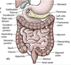
SMALL INTESTINE
3 parts
main function
duodenum
jejunum
ileum
main function:
absorption of nutrients from ingested materials
Key Terms
SMALL INTESTINE
3 parts
main function
duodenum
jejunum
ileum
DUODENUM
first + shortest (25 cm) part
widest + most fixed part
4 parts of the DUODENUM
Superior (first) part:
Short (5 cm); lies anterolateral to bod...
Which part of the duodenum has a mesentery and is mobile?
ampulla (duodenal cap)
first 2 cm of the superior part
superior part of the duodenum ascends from the pylorus and is overlapped by what structures?
liver + gallbladder
(Peritoneum covers its anterior aspect, but it is bare of peritoneu...
The proximal part of the superior duodenum has what 2 structures attached to it?
- hepatoduodenal ligament (part of the lesser omentum) attached superiorly
Related Flashcard Decks
Study Tips
- Press F to enter focus mode for distraction-free studying
- Review cards regularly to improve retention
- Try to recall the answer before flipping the card
- Share this deck with friends to study together
| Term | Definition |
|---|---|
SMALL INTESTINE
| duodenum jejunum ileum main function: absorption of nutrients from ingested materials |
DUODENUM |
(duodenojejunal flexure = L2 vertebra, 2 - 3 cm L. of midline)
(mostly fixed by peritoneum to structures on post. ab. wall) |
4 parts of the DUODENUM | Superior (first) part:
Descending (second) part:
Horizontal (third) part:
Ascending (fourth) part:
|
Which part of the duodenum has a mesentery and is mobile? | ampulla (duodenal cap)
(distal 3 cm of the superior part are immobile –> retroperitoneal) |
superior part of the duodenum ascends from the pylorus and is overlapped by what structures? | liver + gallbladder (Peritoneum covers its anterior aspect, but it is bare of peritoneum posteriorly, except for the ampulla) |
The proximal part of the superior duodenum has what 2 structures attached to it? | - hepatoduodenal ligament (part of the lesser omentum) attached superiorly
|
The bile and main pancreatic ducts enter the posteromedial wall of whiche duodenal section? | DESCENDING (lies to right of/parallel to the IVC; curves inferiorly aroudn head of pancreas) |
hepatopancreatic ampulla |
|
DESCENDING DUODENUM |
|
HORIZONTAL DUODENUM |
|
ASCENDING DUODENUM |
|
Ligament of Treitz |
|
arteries of the duodenum | arise from celiac trunk + superior mesenteric artery celiac trunk:
Superior Mesenteric Artery:
|
pancreaticoduodenal arteries |
–THIS IS THE JUNCTION OF THE EMBRYOLOGICAL FORGUT/MIDGUT–
|
veins of the duodenum |
|
lymphatic vessels of the duodenum |
- anterior lymphatic vessels: drain into the pancreaticoduodenal lymph nodes (along superior + inferior pancreaticoduodenal arteries) + into pyloric lymph nodes (along gastroduodenal artery) - posterior lymphatic vessels: pass posterior to the head of the pancreas, drain into superior mesenteric lymph nodes - Efferent lymphatic vessels: drain into celiac lymph nodes |
nerves of the duodenum |
|
Duodenal (peptic) ulcers | inflammatory erosions of the duodenal mucosa
(ex. erosion of the gastroduodenal artery = severe hemorrhage +peritonitis) |
Which structures may become adherent to the inflamed duodenum and also become ulcerated as the lesion continues to the tissue that surrounds it? (due to close proximity) | liver gallbladder pancreas |
Paraduodenal Hernias |
If a loop of intestine enters this fossa, it may strangulate
|