Anatomy: The Abdomen I
This set of flashcards outlines the anatomical structure and divisions of the abdomen, including its physical boundaries, functional significance, anatomical planes used for medical and anatomical reference, and the nine standard abdominal regions. It also details key landmarks used to define the sagittal and transverse planes.
Abdomen = part of the trunk between the thorax + pelvis.
What are its superior, inferior, anterolateral borders?
superior = diaphragm
inferior = muscles of the pelvis
anterolaterally = musculoaponeurotic walls
Key Terms
Abdomen = part of the trunk between the thorax + pelvis.
What are its superior, inferior, anterolateral borders?
superior = diaphragm
inferior = muscles of the pelvis
...
What are 2 principle functions of the abdomen?
protect + enclose abdominal contents
flexibility = fo...
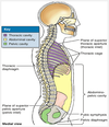
Name the 2 transverse planes + 2 sagittal planes of the abdomen
Transverse:
subcostal plane (most superior)
Name the 9 regions of the abdomin, as numbered in this figure.
Right hypochondriac
Right lumbar
What are the superior and inferior landmarks for the 2 sagittal planes ?
Superior = midclavicular planes; pass approximately 9 cm from the midline
What are the landmarks for the 2 transverse abdominal planes?
subcostal plane = passes through inferior border of the 10th costal cartilage on each side
Related Flashcard Decks
Study Tips
- Press F to enter focus mode for distraction-free studying
- Review cards regularly to improve retention
- Try to recall the answer before flipping the card
- Share this deck with friends to study together
| Term | Definition |
|---|---|
Abdomen = part of the trunk between the thorax + pelvis. What are its superior, inferior, anterolateral borders? | superior = diaphragm inferior = muscles of the pelvis anterolaterally = musculoaponeurotic walls |
What are 2 principle functions of the abdomen? | protect + enclose abdominal contents flexibility = for respiration, posture, locomotion |
Name the 2 transverse planes + 2 sagittal planes of the abdomen | Transverse:
Sagittal:
|
Name the 9 regions of the abdomin, as numbered in this figure. |
|
What are the superior and inferior landmarks for the 2 sagittal planes ? | Superior = midclavicular planes; pass approximately 9 cm from the midline inferior = midinguinal points; midway between the anterior superior iliac spine and the superior edge of the pubic symphysis on each side |
What are the landmarks for the 2 transverse abdominal planes? | subcostal plane = passes through inferior border of the 10th costal cartilage on each side transtubercular plane = passes through the iliac tubercles (approx. 5 cm posterior to anterior superior iliac spine) + body of L5 |
Where is the transpyloric plane located? | approximate midpoint between: superior border of manubrium + superior border of pubic symphysis (L1 vertebral level)
|
What other important structures are transected by the transpyloric plane? |
|
Where does the interspinous plane pass through | anterior superior iliac spine on each side (easily palpated) |
Which part of the abdominal wall is NOT musculoaponeurotic? | posterior abdominal wall (includes lumbar vertebral column) |
The anterolateral abdominal wall extends from__________ to ___________? | thoracic cage to the pelvis |
What are the superior and inferior boundaries of the anterolateral abdominal wall? | superior = 7th - 10th rib cartilages + xiphoid process inferior = inguinal ligament + superior margins of the anterolateral aspects of the pelvic girdle (iliac crests, pubic crests, + pubic symphysis). |
Identify the numbered muscles of the anterior abdominal wall: |
|
Identify the fascia in the anterior abdominal wall: |
|
Identify these anterior abdominal wall structures: |
|
In what area is the skin firmly attached to the subcutaneous tissue (in the anterior abdominal wall) ? | at the unbilicus |
Why is it structurally significant that the muscle fibers of the abdominal walls go in 3 different directions? | b/c the abdomin is a positive pressure area
|
The subcutaneous tissue over most of the abdominal walls is a major storage site for what? What happens if there is too much of this substance stored here? | FAT too much fat** = obesity;** can cause sagging folds “panniculi” (apron) |
There are some areas of the body that maintain fat storage even during starvation. These areas include…? |
fun facts from Dr. Ray :) |
|
|
The membranous layer (Scarpa fascia) continues inferiorly into the ___________. | perineal region as the superficial perineal fascia (Colles fascia) NOT into the thighs (fascia lata) |
What is the clinical significance of the membranous Scarpa fascia being sufficiently complete ? | Significant because: fluids** escaping from a ruptured vessel or urethra (blood and/or urine) may **accumulate deep to it |
The superficial, intermediate, and deep layers of investing fascia cover____________ ? | the external aspects of the three muscle layers of the anterolateral abdominal wall + their aponeuroses (flat expanded tendons) |
The endoabdominal fascia (membranous sheet of varying thickness) lines the __________? | the internal aspect of the abdominal wall |
The portion of fascia lining the deep surface of the transverse abdominal muscle and its aponeurosis is called…? | "transversalis fascia" | (relatively firm) |
The portion of fascia lining the abdominal cavity is called…? | "parietal peritoneum" (internal to transversalis fascia; separated by extraperitoneal fat) |
What is the clinical significance of fascia and fascial spaces of abdominal wall? | 1. potential space b/t membranous layer of subcutaneous tissue + deep fascia (covering rectus abdominis + external oblique m.) fluid may accumulate
|
Why is the potential (or fat-filled) space b/t the endoabdominal fascia of special importance in surgery? | it can be opened during surgery
|
How many bilaterally paired muscles are there in the anterolateral abdominal wall? | 5 muscles
|
The fibers of which 3 abdominal muscles are blended together for increased strength? | external oblique internal oblique transverse abdominal |
External Obliques | origin: External surfaces of 5th - 12th ribs insertion: Linea alba, pubic tubercle + anterior half of iliac crest innervation: Thoracoabdominal nerves (T7 - T11) + subcostal nerve
|
The 3 flat muscles continue anteromedially as….? | aponeuroses | (strong, sheet-like) |
At what point do the aponeuroses form the tough, aponeurotic, tendinous rectus sheath? |
|
Linea alba |
|
|
|
External Oblique Aponeurosis | Inferior attachment = pubic crest (medial to pubic tubercle) inferior margin = inguinal ligament (thickened fibrous band; free posterior edge spanning b/t anterior superior iliac spine + pubic tubercle) |
Inguinal Ligament |
(iliopsoas muscle + femoral vessels + nerve) most herniations happen here = weak area |
Internal Oblique Muscle | Origin: Thoracolumbar fascia, anterior two-thirds of iliac crest + lateral half of inguinal ligament Insertion: Inferior borders of 10th - 12th ribs, linea alba + pecten pubis via conjoint tendon Innervation: Thoracoabdominal nerves (anterior rami of T7-T12 nerves) + first lumbar nerves Main action: Compress + support abdominal viscera; flex + rotate trunk
|
Transversus Abdominis muscles | Origin: Internal surfaces of 7th - 12th costal cartilages, thoracolumbar fascia, iliac crest + lateral third of inguinal ligament Insertion: Linea alba w. aponeurosis of internal oblique, pubic crest + pecten pubis via conjoint tendon Innervation: Thoracoabdominal nerves (anterior rami of T7-T12 nerves) first lumbar nerves Main action: Compresses + supports abdominal viscera
|
Rectus Abdominis (characteristics) |
(broad + thin superiorly; narrow + thick inferiorly)
|
The connecting inersection of the rectus abdominis usually occur at which locations? |
|
Rectus Abdominis Muscle | Origin: Pubic symphysis and pubic crest Insertion: Xiphoid process and 5th - 7th costal cartilages Innervation: Thoracoabdominal nerves (anterior rami of T7-T12 nerves) Main action: Flexes trunk (lumbar vertebrae) + compresses abdominal viscera; stabilizes and controls tilt of pelvis (balancing pelvis) |
Pyramidalis muscle | - absent in approximately 20% of people
|
Rectus Sheath |
|
Rectus Sheath: Layers + make-up |
|
Why is the posterior layer of the rectus sheath deficient superior to the costal margin? | because the transverse abdominal muscle pass internal to the costal cartilages and the internal oblique attaches to the costal margin… |
What are the major functions & actions of the anterolateral abdominal muscles? | →Form a strong expandable support for the anterolateral abdominal wall. →Protect the abdominal viscera from injury. →Compress the abdominal contents to maintain or increase the intra-abdominal pressure and, in so doing, oppose the diaphragm (increased intra-abdominal pressure facilitates expulsion). →Move the trunk and help maintain posture. |
What is important to consider when palpating the anterolateral abdominal walls? |
--> sign of abdominal organ inflammation --> involuntary muscular spasms attempt to protect viscera fr. pressure (painful when abdominal infection is present) |
Palpation of abdominal viscera is performed with the patient in what position? Why? | 1. supine position with thighs and knees semiflexed
|
With a patient is supine with ab. muscles relaxed, what motion stimulates the superficial abdominal reflex? | quickly stroking horizontally (lateral to medial) toward the umbilicus
|
What underlies the linea alba at the umbilicus? What was the function of this structure? | umbilical ring
|
Why is a prominent abdomen normal in infants and young children? |
|
What may be a sign of increased intra-abdominal pressure? | Eversion of the umbilicus - usually resulting from ascites (serous fluid accumulation) or a large mass (tumor; enlarged organ) |
Tumors and organomegaly can produce…? | abdominal enlargement |
What is the clinical significance of being able to palpate the spleen? | it means it is already pathology (3x larger than normal) cannot palpate a normal spleen |
Most abdominal hernias occur in what areas? | inguinal region umbilical region epigastric region |
What type of hernia is common in newborns? What causes this? | Umbilical hernias (Herniation occurs through the umbilical ring)
|
Who will be most likely to experience aquired umbilical hernias? What occurs in this type of hernia? | women + obese people
|
Which parts of the abdominal muscles are more likely to herniate? | - the lines where abdominal aponeuroses interlace (ocassionally have gaps; ex. midline, transition fr. aponeurosis to r.sheath)
OR consequences of surgery or trauma |
epigastric hernia | occurs in midline b/t xiphoid process + umbilicus (through linea alba) (usually problems with muscle fibers at this site = hernia) |
Spigelian hernias | occur along semilunar lines
|
What parts of the Thoracoabdominal nerves supply the anterior abdominal wall? | distal, abdominal parts of the anterior rami of T7 - T11 |
Which lateral (thoracic) cutaneous branches supply the anterior abdominal wall? | from the thoracic spinal nerves T7 - T9 or T10 |
Subcostal nerve | large anterior ramus of spinal nerve T12 |
Iliohypogastric and ilioinguinal nerves | terminal branches of the anterior ramus of spinal nerve L1 |
Which nerves approach the abdominal musculature separately to provide the multi-segmental innervation of the abdominal muscles? | inferior thoracic spinal nerves (T7 - T12) + iliohypogastric and ilioinguinal nerves (L1) |
Inferior thoracic spinal nerves (T7 - T12) + the iliohypogastric and ilioinguinal nerves (L1) run oblique but mostly horizontal. What are they susceptable to? | injury in surgical incisions or from trauma at any level of the abdominal wall |
Injury to the inferior thoracic or iliohypogastric and ilioinguinal nerves may result in what? What does this predispose a person to if it occurs in the inguinal region? |
|
When it is possible, how are abdominal inscisions made? What 2 things are considered in these incisions? | Made to follow the cleavage lines (Langer lines) in the skin insicions chosen for:
|
The location of an incision in abdominal surgery depends on…? What is considered to accomodate these desired outcomes? |
surgeon must consider direction of the muscle fibers + location of aponeuroses and nerves |
What are 2 types of high-risk incisions? | pararectus and inguinal incisions |
Pararectus incisions | along the lateral border of the rectus sheath
|
Inguinal incisions | for repairing hernias
|
Incisional Hernia | protrusion of omentum (a fold of peritoneum) or an organ through a surgical incision
|
What factors can predispose a patient to an incisional hernia? |
|
Endoscopic (Minimally Invasive) Surgery | tiny perforations of the abdominal wall allow the entry of remotely operated instruments
|