Anatomy: The Abdomen II - Inguial Region
These flashcards explain the anatomical location and importance of the inguinal region (groin), emphasizing its role as a passageway for abdominal structures and its clinical relevance due to the high incidence of hernias. It highlights the greater prevalence of inguinal hernias in males due to anatomical differences in the inguinal canal related to the spermatic cord.
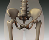
The inguinal region (groin), extends between…
anterior superior iliac spine <—> pubic tubercle
Key Terms
The inguinal region (groin), extends between…
anterior superior iliac spine <—> pubic tubercle
The inguinal region is important anatomically because…
it is a region where structures exit and enter the abdominal cavity
The inguinal region is important clinically because…
the pathways of exit and entrance are potential sites of herniation.
inguinal hernias account for _________% of all abdominal hernias?
75% - 90%
Which gender statistically experiences more inguinal hernias?
What is the percentage and why do this occur?
occur in both sexes, but more commonly in males
approxi...
The inferior migration of which male structre (originally formed in the abdomen) accounts for many of the structural features of the inguinal canal?
the testis
Related Flashcard Decks
Study Tips
- Press F to enter focus mode for distraction-free studying
- Review cards regularly to improve retention
- Try to recall the answer before flipping the card
- Share this deck with friends to study together
| Term | Definition |
|---|---|
The inguinal region (groin), extends between… | anterior superior iliac spine <—> pubic tubercle |
The inguinal region is important anatomically because… | it is a region where structures exit and enter the abdominal cavity |
The inguinal region is important clinically because… | the pathways of exit and entrance are potential sites of herniation. |
inguinal hernias account for _________% of all abdominal hernias? | 75% - 90% |
Which gender statistically experiences more inguinal hernias? What is the percentage and why do this occur? | occur in both sexes, but more commonly in males
because the spermatic cord passes through the inguinal canal; it is bigger than the round ligament in female therfore, the canal is larger making it weaker. |
The inferior migration of which male structre (originally formed in the abdomen) accounts for many of the structural features of the inguinal canal? | the testis |
The inguinal ligament (and iliopubic tract) extends from ……. | anterior superior iliac spine to the pubic tubercle |
The inguinal ligament is a ______________ of the hip joint? | a bilaminar anterior (flexor) retinaculum |
What does the a bilaminar anterior (flexor) retinaculum (inguinal ligament) span, and which structure pass through this space? | -spans the subinguinal space - flexors of the hip and the neurovascular structures serving much of the lower limb pass through the space |
The inguinal ligament is a dense band constituting the inferiormost part of the aponeurosis of which anterior abdominal muscle? | external oblique |
Most of the fibers of the inguinal ligament’s medial end insert into….? | the pubic tubercle |
Some of the deeper fibers of the inguinal ligament pass posteriorly to attach to the ___________? These fibers form the _______________, which forms the medial boundary of the subinguinal space. |
|
The most lateral fibers of the inguinal ligament continue to run along the pecten pubis, forming the ____________? | Pectineal ligament (of Cooper) |
Some of the more superior fibers of the inguinal ligament fan upward, crossing the linea alba + blending with the lower fibers of the contralateral external oblique aponeurosis. These form what ligament? | These fibers form the reflected inguinal ligament |
Iliopubic tract | thickened inferior margin of the transversalis fascia
|
The myopectineal orifice is a region of the groin spanned by what two structures? Is it considered to be strong or weak? | inguinal ligament and iliopubic tract
|
The myopectineal oriface is the site of what common type of injury? What 2 types of this injury are frequently seen here? | groin hernias
|
Describe the inguinal canal in adults |
|
What is the main structure passing through the inguinal canal in males? What is the main structure in females? | males = spermatic cord females = roung ligament
|
Where do the testes develop? | in the extraperitoneal connective tissue in the superior lumbar region of the posterior abdominal wall |
What is the fibrous cord connecting the primordial testis to the anterolateral abdominal wall? This is the future site of what? |
|
What is the processus vaginalis? |
|
1. The testis is in the pelvis by what week of development? 2. Testis lies close to the developing deep inguinal ring by what week? 3A. Testis begins to pass through the inguinal canal during what week? 3B. This process takes approx. how many days? | 1) 12th week of development 2) by 28 weeks (7th month) 3A) during the 28th week; 3B) takes 3 days |
At what time does the testis enter the scrotum? | around month 9 (approx. 4 weeks after passing through the inguinal canal) |
As the testis, its duct (the ductus deferens), and its vessels and nerves descend, they are ensheathed by ____________________? These account for the presence of their derivatives in the adult scrotum: the_____________ and _____________? | - musculofascial extensions of the anterolateral abdominal wall - internal and external spermatic fasciae + cremaster muscle |
The stalk of the processus vaginalis normally degenerates; however, its distal saccular part forms the ______________, the serous sheath of what 2 structures? | "tunica vaginalis testis"
(usually obliterates by the 6th month of fetal development) |
The ovaries develop in the what part of the body? Where do they migrate to? | superior lumbar region of the posterior abdominal wall
|
As in the male, the processus vaginalis of the peritoneum traverses the transversalis fascia at the site of the deep inguinal ring, forming the inguinal canal as in females. What structure does it protrude into? | protrudes into the developing labium majus |
What structures are connected by the female gubernaculum? | ovary and primordial uterus is connected to the developing labium majus |
After birth, the female gubernaculum is represented by what structure? (2 different names in 2 different areas) | ovarian ligament (b/t ovary + uterus) round ligament of uterus (b/t uterus and labium majus) * (image is premature) |
Because of the attachment of the ovarian ligaments to the uterus, the ovaries do not descend to the inguinal region; however, the round ligament passes through the inguinal canal and attaches to the ________________? | subcutaneous tissue of the labium majus |
The inguinal canals in females are wider/narrower than those in males? The canals in infants of both sexes are shorter/longer and much less oblique than in adults? |
|
Where do superficial inguinal rings in infants lie? | almost directly anterior to the deep inguinal rings |
What is the internal entrance to the inguinal canal called? Where is it located? | "deep (internal) inguinal ring" located:
*it is the beginning of an ivagination in the transversalis fascia = forms an openi |
The transversalis fascia itself continues into the inguinal canal, forming_____________? | innermost covering (internal fascia) of structures that traverse the canal |
What is the inferior opening of the inguinal canal called? | superficial (external) inguinal ring |
What are crura? Where do the lateral crus and medial crus attach to? | parts of the external oblique aponeurosis that lie lateral and medial to, and form the margins of, the superficial ring lateral crus = pubic tuberacle medial crus = pubic crest |
intercrural fibers |
|
Between its 2 openings, the inguinal canal has two walls (anterior and posterior), as well as a roof and floor. What is its normal position? | collapsed anteroposteriorly against the structures it conveys |
What are the boundaries of the following inguinal canal regions?
| 1. Anterior wall: aponeuroses of the external oblique and internal oblique muscles. 2. Posterior wall: aponeurosis of the transverse abdominal muscle and transversalis fascia. 3. Superior wall (roof): arching fibers of the internal oblique and transverse muscles. 4. Inferior wall (floor): inguinal and lacunar ligaments. |
Why don't the deep and superficial inguinal rings in the adult overlap? | because of the oblique path of the inguinal canal |
What effect does intra-abdominal pressure have on the inguinal canal? |
(until the pressures overcome the resistant effect of this mechanism) |
What effect does contraction of the external oblique have on the inguinal canal? |
|
When the muscles forming the lateral part of the arches of the internal oblique and transverse abdominal muscles contract, what effect does it have on the inguinal canal? | it makes the roof of the canal descend, constricting the canal |
Inguinal Hernias | protrusion of parietal peritoneum and viscera, such as the small intestine, through a normal/abnormal opening from the cavity in which they belong
|
Indirect inguinal hernia |
|
Direct inguinal hernia | - occurs directly through a weakened area of the abdominal wall muscles (posterior wall of the inguinal canal), lateral to the edge of the conjoint tendon (falx inguinalis), in the inguinal triangle
|
Predisposing factors for direct (aquired) hernias | weakness of anterior abdominal wall in inguinal triangle owing to:
|
Predisposing factors for indirect (congenital) hernias | patency of processus vaginalis (complete or at least superior part) in younger persons
|
FACT CARD: The peritoneal part of the hernial sac of an indirect inguinal hernia is formed by the persisting processus vaginalis. | FACT CARD: If the entire stalk of the processus vaginalis persists, the hernia extends into the scrotum superior to the testis, forming a complete indirect inguinal hernia. |
Direct (aquired) Hernia:
|
(lies outside inner 1 or 2 fascial coverings)
EXITS THRU: superficial ring, lateral to cord. (rarely enters scrotum) |
Indirect (congenital) Hernia:
|
EXITS THRU: superficial ring inside cord (commonly passing in to scrotum/labium majus) |