Understanding Human Anatomy Cells, Tissues, and Or

Learn levels of biological hierarchy, cell structures, tissue types, organ systems, and cell division (Mitosis phases). Perfect for TEAS, nursing, or medical prep.

Mason Bennett
Contributor
4.0
34
4 months ago
Preview (12 of 39 Pages)
100%
Purchase to unlock

Page 1

Understanding Human Anatomy Cells, Tissues, and Or - Page 1 preview image

Loading page image...

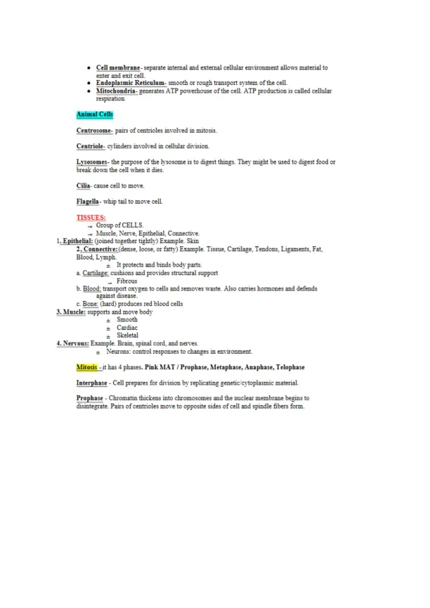
ANATOMY & PHYSIOLOGYAnatomy:is what you see with your eyes in the human body.Microscopic Anatomy:examines cells and molecules.±Cytology:studyofcells.±Histology:study of tissues.Physiology:is the study of functions of anatomical structures.*Smallest living is aCELL.*Smallest organisms is aATOM.Levels of HierarchyAtom-the most basic complete uni: of an element.Molecule-a group of atoms bonded together, representing the smallest fundamental unit of achemical compound that can take part in a chemical reaction.Organelles-are cells parts that function within a cell.Cells- the basic structural unit of an organism from which living things created. Is one individualcell.Tissues- a group of cells with similar structure that functions together as a unit, but at a lowerlevel than organs.Organ-a self contained part of an organism that performs specific functions. Is formed by twoor more similar tissues.Organ System-functionalgroups of organs that work together withinthebody:circulatory,integumentary, skeletal, reproductive, digestive, urinary, respiratory, endocrine, lymphatic,muscular and nervous.±Humans have11Organ Systems.CellsStructureNucleus-holds the cells DNA in form of chromatinRibosomes- small structures that build proteins "amino acids".GolgiApparatus- modifies and packages proteins secreted from cell.Vacuoles-storage, digestion and waste removal.Cytoskeletal-series of rod shaped proteins that provide shape support cell.Microtubules-part of the cytoskeletal.Cytosol-liquid material in cell.

Page 2

Understanding Human Anatomy Cells, Tissues, and Or - Page 2 preview image

Loading page image...

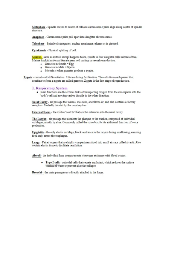
*Cell mem brane-separate internal and external cellular environment allows materialtoenter and exit cell.*EndopJasmicyReticuluin-smooth or rough transport system of the cell.*Mitochondria -generates ATP powerhouse of the cell. ATP production is called cellularrespirationAnimal CellsCentrosome- pairs of centrioles involved in mitosis.Centriole- cylinders involved in cellular division.Lysosomes-the purpose of the lysosome is to digest things. They might be used to digest food orbreak down the cell when it dies.Cilia-cause cell to move.Flagella- whip tail to move cell.TISSUES:_Group of CELLS.-* Muscle, Nerve, Epithelial. Connective.1.Epithelial:(joined together tightly) Example. Skin2. Connective: (dense, loose, or fatty') Example. Tissue, Cartilage, Tendons, Ligaments, Fat,Blood. Lymph.=It protects and binds body parts.a. Cartilage: cushions and provides structuralsupport_Fibrousb. Blood: transport oxygen to cells and removes waste. Also carries hormones and defendsagainst disease.c. Bone: (hard) produces red blood cells3.Muscle: supports and move body±Smooth±Cardiac_Skeletal4. Nervous: Example. Brain, spinal cord, and nerves.±Neurons: control responsestochanges in environment.Mitosis- it has4phases.Pink MAT / Prophase. Metaphase, Anaphase, TelophaseInterphase -Cell prepares for division by replicating genetic cytoplasmic material.Prophase -Chromatin thickens into chromosomes and the nuclear membrane begins todisintegrate. Pairs of centrioles move to opposite sides of cell and spindle fibers form.

Page 3

Understanding Human Anatomy Cells, Tissues, and Or - Page 3 preview image

Loading page image...

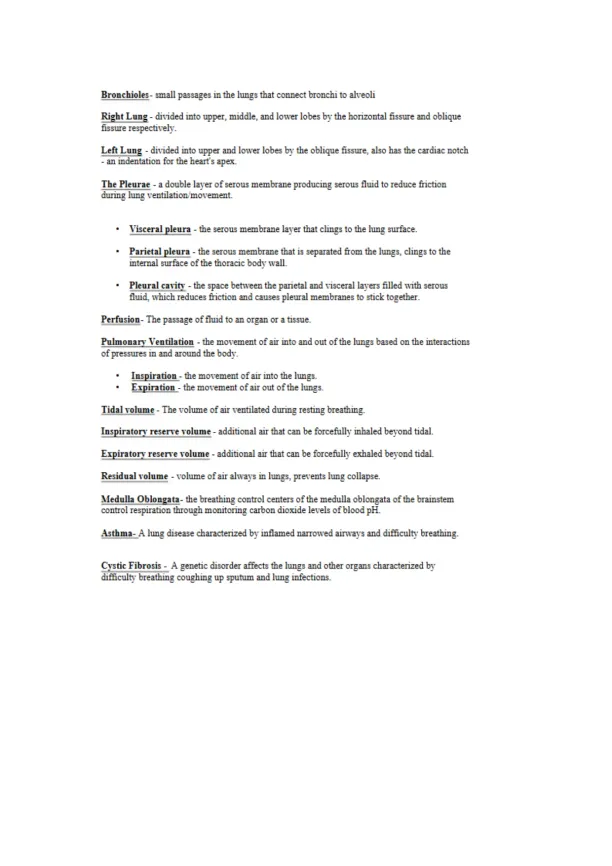
Metaphase -Spindle moves to center of cell and chromosome pairs align along center of spindlestructure.Anaphase -Chromosome pairs pull apart into daughter chromosomes.Telophase -Spindle disintegrates, nuclear membrane reforms or is pinched.Cytokinesis - Physical splitting of cell.Meiosis- same as mitosis except happens tivice, results in four daughter cells instead of two.Mature haploid male and female germ cell uniting in sexual reproduction.±Gametes in female = Egg=Gametes in Male = Sperm=Meiosis is when gametes produce a zygote.Zygote:controls cell differentiation. It forms during fertilization. The cells from each parent thatcombine to form a zygote are called gametes. Zygote is the first stage of reproduction.L Respiratory System«main functions are the critical tasks of transporting oxygen from the atmosphere into thebody's cell and moving carbon dioxide in the other direction.Nasal Cavity -air passage that warms, moistens, and filters air. and also contains olfactoryreceptors. Medially divided by the nasal septum.External Nares - the visible 'nostrils' that are the entrances into the nasal cavityThe Larynx -air passage that connects the pharynx to the trachea, composed of individualcartilages, mostly hyaline. Commonly called the voice box for its additional function of voiceproduction.Epiglottis -the only elastic cartilage, blocks entrance to the lannx during swallowing, ensuringfood only enters the esophagus.Lungs - Paired organs that are highly compartmentalized into small air sacs called alveoli. Alsocontain elastic tissue to facilitate ventilation.Alveoli -the individual lung compartments where gas exchange with blood occurs.TyPe2cells -cuboidal cells that secrete surfactant, which reduces the surfacetension of water to prevent alveolar collapse.Bronchi - the main passageways directly attached to the lungs.

Page 4

Understanding Human Anatomy Cells, Tissues, and Or - Page 4 preview image

Loading page image...

Bronchioles- small passages in the lungs that connect bronchi to alveoliRight Lung -divided into upper, middle, and lower lobes by the horizontal fissure and obliquefissure respectively.Left Lung -divided into upper and lower lobes by the oblique fissure, also has the cardiac notch- an indentation for the heart’s apex.ThePleurae -a double layer of serous membrane producing serous fluid to reduce frictionduring lung ventilation movement.Visceral pleura -the serous membrane layer that clings to the lung surface.Parietal pleura -the serous membrane that is separated from the lungs, clings to theinternal surface of the thoracic body wall.Pleural cavity -the space between the parietal and visceral layers filled uith serousfluid, which reduces friction and causes pleural membranes to stick together.Perfusion - The passage of fluid to an organ or a tissue.PulmonaryVentilation- the movement of air into and out of the lungs based on the interactionsof pressures in and around the body.Inspiration -the movement of air into the lungs.Expiration -the movement of air out of the lungs.Tidal volume - The volume of air ventilated during resting breathing.Inspiratoryreservevolume -additional air that can be forcefully inhaled beyond tidal.Expiratory reserve volume -additional air that can be forcefully exhaled beyond tidal.Residua] volume - volume of air always in lungs, prevents lung collapse.Medulla Oblongata-the breathing control centers of the medulla oblongata of the brainstemcontrol respiration through monitoring carbon dioxide levels of blood pH.Asthma- A lung disease characterized by inflamed narrowed airways and difficulty breathing.Cystic Fibrosis -A genetic disorder affects the lungs and other organs characterized bydifficulty breathing coughing up sputum and lung infections.

Page 5

Understanding Human Anatomy Cells, Tissues, and Or - Page 5 preview image

Loading page image...

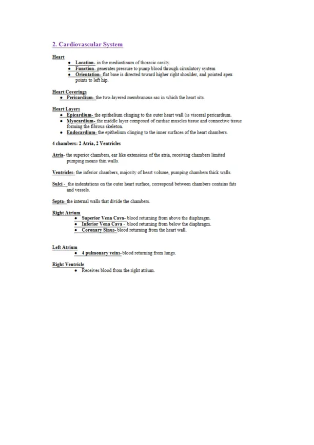
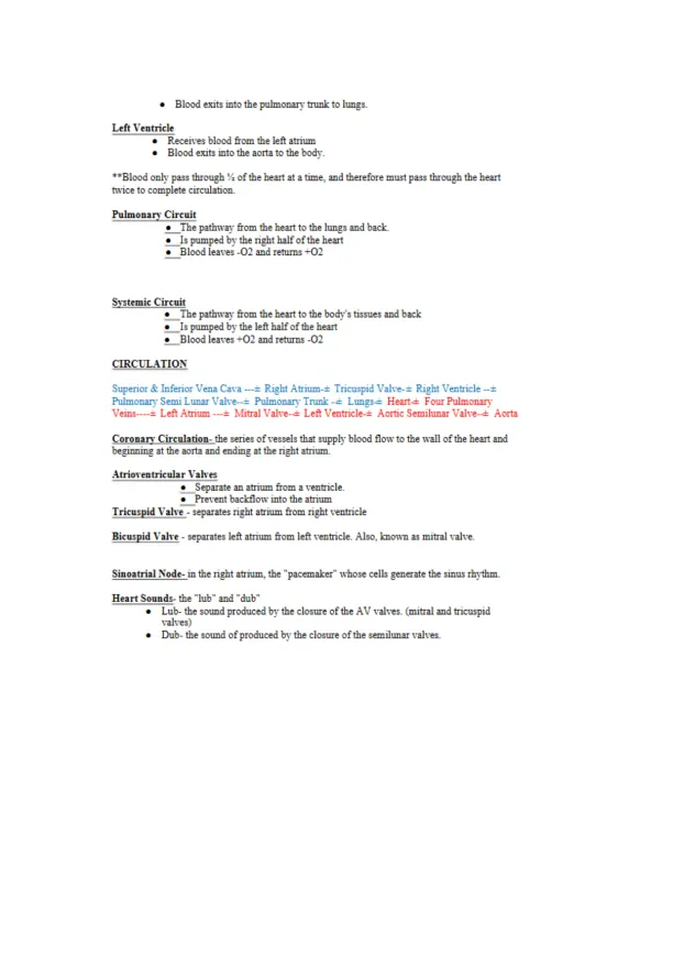
2. Cardiovascular SvstemHeartLocation-in the mediastinum of thoracic cavity.Function-generates pressure to pump blood through circulator}’ systemOrientationflat base is directed toward higher right shoulder, and pointed apexpoints to left hip.Heart Coverings*Pericardium-the two-layered membranous sac in which the heart sits.Heart LayersEpicardium-the epithelium clinging to the outer heart wall (is visceral pericardium.*Myocardium-the middle layer composed of cardiac muscles tissue and connective tissueforming the fibrous skeleton.Endocardium-the epithelium clinging to the inner surfaces of the heart chambers.4 chambers: 2 Atria, 2 VentriclesAtria-the superior chambers, ear like extensions of the atria, receiving chambers limitedpumping means thin walls.Ventricles- the inferior chambers, majority of heart volume, pumping chambers thick walls.Sulci - the indentations on the outer heart surface, correspond between chambers contains fatsand vessels.Septa-the internal walls that divide the chambers.Right AtriumSuperiorVenaCava-blood returning from above the diaphragm.Inferior Vena Cava -blood returning from below the diaphragm.Coronary Sinus-blood returning from the heart wall.Left Atrium4 pulmonary veins-blood returning from lungs.Right VentricleReceives blood from the right atrium.

Page 6

Understanding Human Anatomy Cells, Tissues, and Or - Page 6 preview image

Loading page image...

Blood exits into the pulmonary trunk to lungs.Left VentricleReceives blood from the left atriumBlood exits into the aorta to the body.**Blood only pass through % of the heart at a time, and therefore must pass through the hearttwice to complete circulation.PulmonaryCircuit_The pathway from the heart to the lungs and back.Is pumped by the right half of the heartBlood leaves -02 and returns +02Systemic CircuitThe pathway from the heart to the body's tissues and backIs pumped by the left half of the heartBlood leaves +02 and returns -02CIRCULATIONSuperior & Inferior Vena Cava —± Right Atrium-± Tricuspid Valve-± Right Ventricle —±Pulmonary Semi Lunar Valve—± Pulmonary Trunk --= Lungs-= Heart-= Four PulmonaryVeins— ± Left Atrium —= Mitral Valve--= Left Ventricle-= Aortic Semilunar Valve--± AortaCoronary7Circulation- theseries of vessels that supply blood flow to the wall of the heart andbeginning at the aorta and ending at the right atrium.Atrioventricular ValvesSeparate an atrium from a ventricle.Prevent backflow into the atriumTricuspid Valve- separates right atrium from right ventricleBicuspid Valve- separates left atrium from left ventricle. Also, known as mitral valve.Sinoatrial Node- in the right atrium, the "pacemaker" whose cells generate the sinus rhythm.Heart Sounds- the "lub" and "dub"Lub- the sound produced by the closure of the AV valves, (mitral and tricuspidvalves)Dub- the sound of produced by the closure of the semilunar valves.

Page 7

Understanding Human Anatomy Cells, Tissues, and Or - Page 7 preview image

Loading page image...

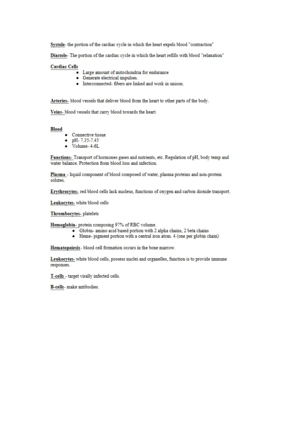
Systole-the portion of the cardiac cycle in which the heart expels blood "contraction"Diastole-The portion of the cardiac cycle in which the heart refills with blood "relaxation'Cardiac CellsLarge amount of mitochondria for enduranceGenerate electrical impulses.Interconnected- fibers are linked and work in unison.Arteries- blood vessels that deliver blood ban the heart to other parts of the bod}-.Veins- blood vessels that cany blood towards the heartBloodConnective tissuepH-7.35-7.45Volume- 4-6LFunctions-Transport of hormones gases and nutrients, etc. Regulation of pH. body temp andwater balance. Protection from blood loss and infection.Plasma - liquid component of blood composed of water, plasma proteins and non-proteinsolutes.Erythrocytes-red blood cells lack nucleus, functions of oxygen and carbon dioxide transport.Leukocytes-white blood cellsThrombocytes- plateletsHemoglobin-protein composing 97% of RBC volume.Globin- amino acid based portion with 2 alpha chains. 2 beta chainsHeme- pigment portion with a centra] iron atom.4(one per globin chain)Hematopoiesis -blood cell formation occurs in the bone marrow.Leukocytes-white blood cells, possess nuclei and organelles, function is to provide immuneresponses.T-cells - target vitally infected cells.B-cells- make antibodies.

Page 8

Understanding Human Anatomy Cells, Tissues, and Or - Page 8 preview image

Loading page image...

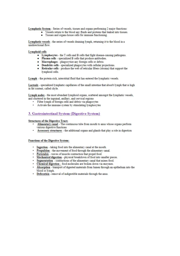
Lymphatic System - Series of vessels, tissues and organs performing 2 major functions:Vessels return to the blood any fluids and proteins that leaked into tissues.Tissues and organs house cells for immune functioning.Lymphatic vessels - the series of vessels draining lymph, returning it to the blood in aunidirectional flow.Lymphoid cellsLymphocytes -the T cells and B cells that fight disease-causing pathogens.Plasmacells- specialized B cells that produce antibodies.Macrophages -phagocytize any foreign cells or debris.Dendritic cells -specialized phagocytes with cellular projections.Reticular cells -produce the web of reticular fibers (stroma) that support thelymphoid cells.Lymph - the protein-rich, interstitial fluid that has entered the lymphatic vessels.Lacteals - specialized lymphatic capillaries of the small intestine that absorb lymph that is highin fat content, called chyle.Lymph nodes - the most abundant lymphoid organs, scattered amongst the lymphatic vessels,and clustered in the inguinal, axillary. and cervical regionsFilter lymph of foreign cells and debris via phagocytesActivate the immune system by stimulating lymphocytes3. Gastrointestinal System (Digestive System)Structures of the Digestive Tract:Alimentary canal - The continuous tube from mouth to anus whose organs performvarious digestive functionsAccessory structures- the additional organs and glands that play a role in digestionFunctions of the Digestive System:Ingestion -taking food into the alimentary canal at the mouth.Propulsion - the movement of food through the alimentary canal.Peristalsis - waves of muscle contraction that propel food.Mechanical digestion -physical breakdown of food into smaller pieces.Segmentation -contractions of the alimentary canal that mixes food.Chemical digestion -food molecules are broken down via enzymes.Absorption - transport of digested materials from lumen through an epithelium into theblood or lymph.Defecation -removal of indigestible materials through the anus.

Page 9

Understanding Human Anatomy Cells, Tissues, and Or - Page 9 preview image

Loading page image...

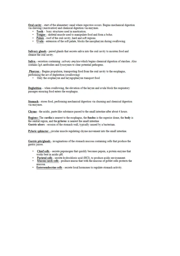
Oralcavity - start of the alimentary canal whereingestionoccurs. Begins mechanical digestionvia chewing(mastication)and chemical digestion via enzymes.Teerh - bony structures used in mastication.Tongue -skeletal muscle used to manipulate food and form a bolus.Palate - roof of the oral cavity, hard and soft regions.Uvula -extension of the soft palate, blocks the nasopharynx during swallowing.Salivary glands -paired glands that secrete saliva into the oral cavity to moisten food andcleanse the oral cavity.Saliva - secretion containing sm'n-aj’yamylasewhich begins chemical digestion of starches. Alsocontains IgA antibodies and lysozymes to clear potential pathogens.Pharynx -Begins propulsion, transporting food from the oral cavity to the esophagus,performing the act of deglutition (swallowing)Only the oropharynx and laryngopharynx transport foodDeglutition -when swallowing, the elevation of the larynx and uvula block the respiratorypassages ensuring food enters the esophagus.Stomach -stores food, performing mechanical digestion via churning and chemical digestionvia enzymes.Chyme - the acidic, paste-like substance passed to the small intestine after about 4 hours.Regions:The cardia is nearest to the esophagus, thefundusis the superior dome, thebodyisthe central region, and thepylorusis nearest the small intestineGastric ulcers- erosion of the stomach wall, typically caused by a bacterium.Pyloric sphincter -circular muscle regulating chyme movement into the small intestine.Gastric pits/glands -invaginations of the stomach mucosa containing cells that produce thegastric juices.Chief cells - secrete pepsinogen that quickly becomes pepsin, a protein enzyme thatworks best in acidic pH.Parietal cells - secrete hydrochloric acid (HCL), to produce acidic environment.Mucous neck cells -produce mucus that with the mucous of goblet cells protects themucosa.Enteroendocrine cells -secrete local hormones to regulate stomach activity.

Page 10

Understanding Human Anatomy Cells, Tissues, and Or - Page 10 preview image

Loading page image...

Small intestine -coiled digestive organ transporting food from stomach to large intestine.Completes digestion and performs all nutrient absorption. Receives secretions form liver, gallbladder, and pancreas.Regions -divided into duodenum, jejunum, and ileumHepatopancreatic sphincter (of Oddi) -regulates the entrance of secretions from the liver, gallbladder and pancreas into the duodenum.Specializations for absorption:*Villi- finger-like projections of foe mucosa that increase surface area of the epithelium.Each villus contains a capillary bed and a lacteal for nutrient absorption.*Intestinal crypts -invaginations producing intestinal juices, a mucous secretion.*Microvilli- finger-like extensions of foe epithelial cell membranes with attached "brushborder enzymes." further increasing surface area.Liver -a 4-lobed gland whose digestive function is the production of bile.Bile - a green, alkaline fluid containing bilirubin pigment and bile salts that performemulsification of fats. Drains from the liver via the hepatic ducts.Emulsification - the physical breakdown of large fat globules into smaller fat droplets, thusincreasing surface area for enzymatic digestion.Gallbladder - muscular sac on the posterior surface of foe liver that stores and concentratesbile. The gall bladder is drained by the cystic duct.ThePancreas -gland whoseacinar ceilsproduces pancreatic juices containing digestiveenzymes such as proteases, lipases, nucleases, and amylases.Large Intestine(Colon) - transports food from foe small intestine to the anus while absorbingwater and forming feces.Deocecal valve - regulates movement of food from the small to the large intestine.Hanstra - individual pouches of the large intestine wall.Teniae coli - thin longitudinal muscle layer forming the haustra.Regions :Cecum - pouch-like region with the hanging vermiform appendix.

Page 11

Understanding Human Anatomy Cells, Tissues, and Or - Page 11 preview image

Loading page image...

*Ascending colon -transports food up the right side of the abdomen, turning at thehepatic flexure.*I ransverse colon- transports food across the abdomen, turning at thesplenic flexure.Descending colon- transports food down the left side of the abdomen.Sigmoid colon- s-shaped region.Rectum -most distal region of the colon. w7here feces is stored until defecation.Recta] valves - projections that allow gases to pass around feces.Anus -endofthe alimentary canal from which defecation occurs.Internal anal sphincter -smooth muscle regulating ana] opening.Eiternalanal sphincter -skeletal muscle regulating anal opening.4. Neuromuscular System (Nervous System)Skeletal Musclej-iber (cells)- long, multinucleated cells containing many proteins and manymitochondria.Sarcolemma-the plasma membrane of a muscle fiber, nuclei just beneath.Sarcoplasm-thecytoplasm of a muscle fiber.Giveosomes-compartments that store glycogen, provides glucose during activity.Myofilaments-the protein-based contractile elements whose specific pattern gives the striatedappearance.Thick Filaments- composed of a bundle of myosin proteins.Thin F:ilaments-composed of actin and other proteins.NMJ-where a motor neurons axon meets a skeletal muscle fiber.ActionPotential-the electrical current that travels along the neuron, then to the sarcolemma totrigger muscle contraction.Acetylcholine (ACh)-neurotransmitter used at NMJ's to trigger contractions, released fromneuron and crosses synaptic cleft.AfferentNerves-sensory nerves that send message to the CNS

Page 12

Understanding Human Anatomy Cells, Tissues, and Or - Page 12 preview image

Loading page image...

Efferent Nen es- motor nerves that send message to muscles.ANS-autonomic nervous system controls involuntary actions including cardiac and smoothmuscle, heart, digestion and breathing.Axon- a nerve fiber.Contraction-elongating or shortening of muscle to perform muscle actions.Nerve- a bundle of axons that transmit electrical impulses to peripheral organs.Synapse-the structure that allows neurons to pass signals to other neurons.5. Reproductive SystemMale ReproductiveTestes- ±e primary sex organs, producing spenn and sex hormones.Sustain developing spenn, and produce proteins used in sperm production.Scrotum-the sac of skin that contains the testes.Provides ideal temp for spermDarios Muscle- contracts to fold scrotum wall.Cremaster Muscle-contracts to elevate testes.Prostate & Seminal Vesicles- produce the fluids necessary for lubricating and nourishingspenn.Penis-the male copulatory system.Testosterone-the hormone that stimulates male secondary sexual characteristics.TheDuct SystemEpididymis -coiled tubules clinging to the testes where sperm mature and gain theirability to swim.Vas deferens- tubule that transports sperm from the epididymis to the prostate gland.Urethra- passage that transports semen and urine.Female ReproductiveOvaries-primary sex organ, producing eggs and sex hormones. Estrogen is produced in ovaries.Fallopian tubes-transports an egg from and ovary to uterusFimbriae-finger-like projections at the opening of the fallopian tube that sweep eggs in.
Preview Mode

This document has 39 pages. Sign in to access the full document!

Study Now!

XY-Copilot AI
Unlimited Access
Secure Payment
Instant Access
24/7 Support
Document Chat

Document Details

Related Documents

View all